Members Login


Channels
Special Offers & Promotions
The solution to high quality fluorescent image deconvolution
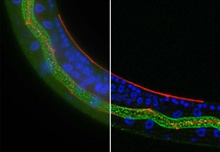 Olympus' new BX3 microscope systems are designed with the
user in mind to maximise the quality of fluorescence images captured as part of
life science research projects. The high-end BX63 system comfortably handles day-to-day
applications, while boasting a range of advanced features to satisfy the most
complex of needs, including rapid deconvolution of fluorescent Z-stack images.
This approach is particularly useful for imaging thicker samples at high
magnification, such as tissues and cells. Deconvolution removes the
out-of-focus blur from each image in the Z-stack to improve clarity and increase
the precision of downstream analyses. This can only be accurately achieved if
the distance between the Z-stack images is stringently controlled. Therefore,
the fully-automated BX63 frame employs a high-precision focusing mechanism adjusted
using the nosepiece, rather than the stage, which increases system stability
and maximises Z-resolution. When combined with the cellSens software suite and
deconvolution solution module, the rapid creation of extremely accurate
deconvolved fluorescent images is only a few clicks away.
Olympus' new BX3 microscope systems are designed with the
user in mind to maximise the quality of fluorescence images captured as part of
life science research projects. The high-end BX63 system comfortably handles day-to-day
applications, while boasting a range of advanced features to satisfy the most
complex of needs, including rapid deconvolution of fluorescent Z-stack images.
This approach is particularly useful for imaging thicker samples at high
magnification, such as tissues and cells. Deconvolution removes the
out-of-focus blur from each image in the Z-stack to improve clarity and increase
the precision of downstream analyses. This can only be accurately achieved if
the distance between the Z-stack images is stringently controlled. Therefore,
the fully-automated BX63 frame employs a high-precision focusing mechanism adjusted
using the nosepiece, rather than the stage, which increases system stability
and maximises Z-resolution. When combined with the cellSens software suite and
deconvolution solution module, the rapid creation of extremely accurate
deconvolved fluorescent images is only a few clicks away. Image deconvolution is a powerful tool in life science research that removes out-of-focus blur and allows the user to create images in which every detail is clearly resolved. This technique is particularly useful when investigating cellular organelles or thick samples at high magnification, as these conditions reduce the depth of field and exacerbate the blurring effect of out-of-focus light. To achieve this without the use of a confocal microscope requires accurate Z-position information, high frame stability, quality optics and complex software algorithms. For this reason, the BX63 frame is fully automated to facilitate extremely precise control of sample movement and focal position. In addition, the microscope is uniquely focused using the nosepiece, allowing the stage to remain fixed in position, thereby increasing stability and accuracy.
To make deconvolution a powerful tool for use in life science research, it needs to be fast and easy to carry out. Using the cellSens software suite, the BX63 can be intuitively controlled making it simple to plan and perform experiments. The Deconvolution Solution module creates sharp images that allow researchers to extract as much information as possible from an experiment, including structures, iso-surfaces and projections (via the included Voxel Viewer).
For more information visit www.microscopy.olympus.eu
Subscribe to any of our newsletters for the latest on new laboratory products, industry news, case studies and much more!

Popular this Month
Top 10 most popular articles this month
Today's Picks
Looking for a Supplier?
Search by company or by product
Please note Lab Bulletin does not sell, supply any of the products featured on this website. If you have an enquiry, please use the contact form below the article or company profile and we will send your request to the supplier so that they can contact you directly.
Lab Bulletin is published by newleaf marketing communications ltd.
Media Partners