With years of experience in the pharmaceutical and life science sector, Creative Diagnostics launches NanoHollows, which contain cavity enclosed by a non-porous nanoshell. NanoHollows have great potential applications in biological sensing, biomedical imaging and photothermal therapy due to their unique localized surface plasmon resonance (LSPR) feature...
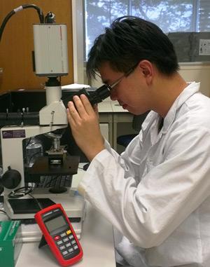
Merck, a leading science and technology company, has launched the CellASIC® ONIX2 Microfluidic System for advanced live cell imaging.
The system converts laboratory microscopes into powerful tools for live cell imaging to more effectively perform in-depth analysis of cellular mechanisms and behavior in a live environment....
DELMIC develops and manufactures products which are focused on high performance, user friendly, integrated microscopy solutions.
Users of Delmic's SECOM solution for Correlated Light & Electron Microscopy (CLEM) have recently published a review in Nature Methods that illustrates the power of CLEM in the world of biology. The boom in the use of super-resolution microscopies in recent years reached a peak with the award of the 2014 Nobel Prize in Chemistry to Betzig, Hell and Moerner for their contributions in the development of super-resolved fluorescence microscopy...
The application of morphologically directed Raman spectroscopy to characterizing the component-specific particulate properties of the active components in a topical pharmaceutical cream is described in an application note from Malvern Instruments.
While it is relatively straightforward to determine the particle size distributions of the individual components of a drug formulation before they are formulated as a blend, doing so post-blending is more challenging. Now, the Morphologi G3-ID, which combines automated image analysis with Raman spectroscopy in a single instrument, enables component-specific particle size and shape distributions to be obtained from a blend. The results of this analysis can provide....
Read MoreXEI Scientific reports a novel approach to removing silicone-based contamination in a recent publication in JVSTDec 2, 2013
XEI Scientific Inc, manufacturer of the popular EVACTRON® De-Contaminator™ Plasma Cleaning System for electron microscopes and other vacuum chambers, is pleased to announce the publication of a paper in collaboration with General Electric's Global Research Center on the use of in-situ plasma cleaning.
The paper appears in the Journal of Vacuum Science & Technology. A contamination, even at extremely low levels, can often hide or distort analyses of surfaces that researchers would like to study. Such is the case of many of the samples analysed at General Electric's Global Research Center in New York. Attempts to study "as received" samples by time of flight secondary ion mass spectrometry (ToF-SIMS) reveal a contamination signature that has come from processing, handling and/or a specific exposure...
Anasys Instruments reports on EPFL's publication in Plant Cell on the use of nanoIR to look into the process of photosynthesis to shed more light on how plants produce energy
École Polytechnique Federale de Lausanne, better known as EPFL, has recently reported on how a group of its scientists have used powerful imaging techniques including nanoIR to support a study which sheds light on photosynthesis. All plants use a form of photosynthesis to produce energy, though not all rely exclusively on it. In higher plants, capturing light takes place in specialized compartments called thylakoids. These are found in cell organelles called chloroplasts, which are the equivalent of a power station for the plant....
Multi-mode microplate readers are an indispensable laboratory instrument that enable many applications to be performed on multiple samples.
Life science applications involving cells are increasingly desired due to higher biological relevance associated with functional readouts. Microplate readers can provide quantification of a specific target, such as a protein or its post-translational modification, or phenotypes such as cell death through apoptosis or necrosis. Quantification is provided relative to assay controls and/or standards using dispersive optical detection and light collection with photomultiplier tubes (PMT)...
Market leaders in temperature controlled microscopy, Linkam Scientific Instruments report on the use of their popular THMS600 stage for polymer research at the Institute of Chemistry and Technology of Polymers (CNR), Italy.
A group of scientists from the CNR is investigating the melting kinetics of poly(3-hydroxybutyrate) (PHB), a natural thermoplastic polymer with mechanical properties comparable to synthetic polymers. PHB is now the focus of scientific interest as it has commercial potential as a fully biodegradable and biocompatible product. Potential uses include environmentally friendly packaging and films, pharmaceutical drug administration, and biocompatible resorbable medical implants. Small batches of PHB have been produced since 1925 by bacterial fermentation, but until now there is no large scale commercial production because it is more expensive than commonly used polymers...
With the introduction of the Morphologi G3-ID system last year, Malvern Instruments is providing researchers with the ability to apply morphologically directed Raman spectroscopy to a range of different sample types
Such advanced particle analysis is proving valuable and highly effective in applications that include inhaled product characterization and pharmaceutical bioequivalence studies, bioequivalence for nasal sprays being referenced specifically in a talk given by the FDA at this year’s IFPAC conference [January 22-25, 2013 Baltimore, MD, USA]. The use of morphologically targeted Raman spectroscopy has also been described in an independent article, as part of granulation studies, to provide the physical and chemical identities of particles in mixed populations [1]...
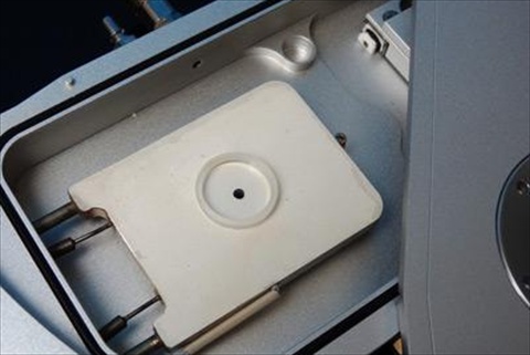
Read MoreXEI Scientific reports on how RJ Lee Group has applied Evactron plasma technology for specimen cleaningJan 15, 2013
XEI Scientific Inc, maker of the popular EVACTRON® De-Contaminator™ Plasma Cleaning System for electron microscopes and other vacuum chambers, reports on how leading contract materials characterization laboratory, RJ Lee Group, Inc., have used an Evactron® specimen cleaning chamber to clean samples ahead of analysis in electron microscopes
RJ Lee Group, Inc. is a materials characterization laboratory and industrial forensics consulting firm employing more than 300 scientists, engineers, technicians and support staff at locations in multiple locations across the USA. The firm specializes in scientific support of four areas of interest: analytical laboratory testing, industrial forensics consultation and support, litigation support, and systems reliability tools for engine health....
Read MoreOcean Thin Films Announces Multispectral Imaging Research Grant Program Nov 9, 2012
Academic, industry professionals can use free or subsidized SpectroCam for data collection
Ocean Thin Films is calling for grant proposals featuring multispectral imaging applications that would utilize the SpectroCam™ platform. SpectroCam is an imaging system that integrates scientific-grade sensors with eight filters to produce multispectral images. This grant gives participants the unique opportunity to add a high-end instrument to their lab at little or no cost...
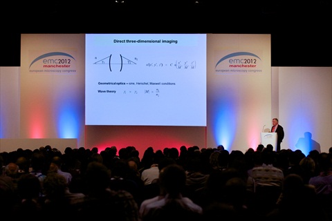
Read MoreRecord number of delegates and visitors flock to the 15th European Microscopy CongressOct 5, 2012
The 15th European Microscopy Congress – emc2012 - held at Manchester Central in September, was the largest yet in the series. It attracted 1,714 registered Conference Delegates. This is a 30% increase on 2008
The record breaking numbers were drawn by eight parallel conference sessions that embraced the life and physical sciences, and delivered a balanced programme of optical and electron microscopy. This breadth made emc2012 the most inclusive event yet. Professor Tony Wilson, President of the Royal Microscopical Society and Vice Chair of the Congress, said, “The conference sessions have provided an unparalleled opportunity for delegates to immerse themselves in their own area of interest, and also to witness new techniques and tools that might benefit their current work, or feature in their future activities.”...
Read MoreReverse Engineering of polymers using nanoscale IR spectroscopy via AFM-IR Aug 6, 2012A recent publication in Spectroscopy Europe by lead authors from Kimberly Clark Corporation showed the use of AFM based nanoscale IR Spectroscopy for reverse engineering of polymeric multilayer films.
The Atomic Force Microscope (AFM) is a fairly common nanoscale characterization technique but its main drawback to date was its inability to provide chemical composition information from a sample. By combining the AFM with a tunable Infrared (IR) source, IR spectra with nanoscale spatial resolution can be collected. As film thicknesses in multilayer films continue to shrink, AFM-IR provides an important capability for sample analysis...
Read MoreSteady Funding Initiatives are Instrumental in Channeling Optical Imaging into New Application Areas, States Frost & SullivanAug 1, 2012Application scope set to expand rapidly from current base of ophthalmology, cardiology, neurology and gastroenterology
Current optical imaging technologies provide reproducible, accurate, objective, quantitative assessments of tissue structures. As the marked progress in optical imaging techniques observed in recent years is instigating a new wave of business opportunities, there are still areas to improve...
Read MoreSteady Funding Initiatives are Instrumental in Channeling Optical Imaging into New Application Areas, States Frost & SullivanJul 16, 2012Application scope set to expand rapidly from current base of ophthalmology, cardiology, neurology and gastroenterology
Current optical imaging technologies provide reproducible, accurate, objective, quantitative assessments of tissue structures. As the marked progress in optical imaging techniques observed in recent years is instigating a new wave of business opportunities, there are still areas to improve. Analysis from Frost & Sullivan's Emerging Trends in Optical Imaging Techniques for Drug Discovery, Clinical Diagnostics and Molecular Imaging research finds that optical imaging at both the macroscopic and microscopic levels is being used intensively by clinicians for diagnosis and treatment-specific applications....
Read MoreThe Science and Engineering Institutes, Singapore, are using Nanoparticle Tracking Analysis to characterize exosomes.Jul 10, 2012NanoSight, leading manufacturers of unique nanoparticle characterization technology, reports on the work of Dr Seow Yiqi at SCEI, Singapore where they apply Nanoparticle Tracking Analysis (NTA) to characterize exosomes for use in delivering therapeutic moieties to specific tissues in vivo.
The Molecular Engineering Laboratory was established in 2009 by Nobel Laureate Dr Sydney Brenner as a multi-disciplinary laboratory where diverse expertise from different realms of science could come together to develop novel technologies. Post-doctoral fellows in the laboratory undertake a wide variety of research ranging from organic fluorophore development to characterization of marine biomaterials with unique physical properties to genetic therapies.
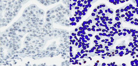
Read MoreLeica Microsystems and Indica Labs Announce Availability of Integrated Advanced Digital Pathology Image AnalysisJul 6, 2012Leica Microsystems, world-leading provider of Total Digital Pathology solutions, and Indica Labs, developing excellence in image analysis for Digital Pathology, today announce the integration of Indica Labs' advanced image analysis algorithm portfolio into Leica Microsystems digital pathology solution.
Users of the Leica Microsystems Tissue IA product can now source from Indica Labs, a range of image analysis algorithms, providing solutions to many diverse tissue-based quantification applications. Indica Labs' image analysis algorithms for molecular assay and morphological feature detection and quantification in whole slide images, coupled with Leica Microsystems' Tissue IA, provides an easy-to-use solution for complex image analysis problems in Digital Pathology...
Read MoreUnderstanding protein crystallization growth at the University of Leeds using a temperature stage from Linkam Scientific InstrumentsJul 3, 2012Market leaders in temperature controlled microscopy, Linkam Scientific Instruments report on the use of their LTS350 stage at the University of Leeds to visualise and grow HEW lysozyme crystals.
The Institute for Particle Science and Engineering is a centre of excellence within the School of Process, Environmental and Materials Engineering at the University of Leeds. Researchers here focus on projects that address the engineering science of particulate processes and products. PhD and MSc students have been using an LTS350 hot-stage system in their research for several years... Read MoreEuropean Microscopy Congress Jun 29, 2012The Scientific Programme for emc2012 comprises eight parallel sessions with the Life Sciences featuring more strongly than ever before.
The Scientific Programme for the 15th European Microscopy Congress includes six plenary talks, 123 invited speakers and 330 oral presentations spread across 72 sessions that embrace light and electron microscopy. The Congress has built its reputation on the Physical Sciences, but recent conferences have included sessions on the Life Sciences... Read MoreNeed for Reduced Healthcare Outlays Creates Strong Growth Potential for Medical Imaging Services, Says Frost & SullivanJun 29, 2012Global Vision with Local Focus will be Key Strategy to Succeed in Highly Fragmented European Medical Imaging Market
The European medical imaging services market is highly fragmented, intensely competitive and beset with pricing pressures. The expansion of service portfolios to include new, customised service structures will be critical to keeping in sync with evolving consumer needs. The most important challenge for market participants will be to conflate a global view with a local approach that addresses specific country market requirements...
Read MoreFrost & Sullivan: Greater Public Funding, Paralleled by Increasing Profile of Private Healthcare, to Spur Medical Imaging Market in CEE and RussiaMay 29, 2012Provision of Alternative Financing by Medical Imaging Vendors and Governmental Support for Public-private Partnerships to Accelerate Market Growth
Rising disease incidence and growing awareness about health issues will boost demand for medical imaging services across Central and Eastern Europe (CEE). However, the installed base of imaging devices in the region is insufficient, especially in the high-end segment, such as magnetic resonance imaging (MRI), computed tomography (CT) and positron emission tomography (PET). At the same time, a large proportion of lower-end imaging modalities is obsolete and requires replacement. The availability of EU funds and governmental financial support, as well as the increasing importance of private healthcare, are set to help overcome this challenge.
Read MoreAgar Scientific announces new highly concentrated colloidal gold for research applicationsMay 17, 2012Agar Scientific, a leading supplier of microscopy accessories and consumables, announces a new range of highly concentrated gold colloidal nanoparticles from British Biocell International (BBI). Agar Scientific is a market leader in the supply of high quality accessories to assist with sample handling for the microscopy market serving a very broad range of application areas. One of these ranges is the gold nanoparticles produced by BBI...
Read More

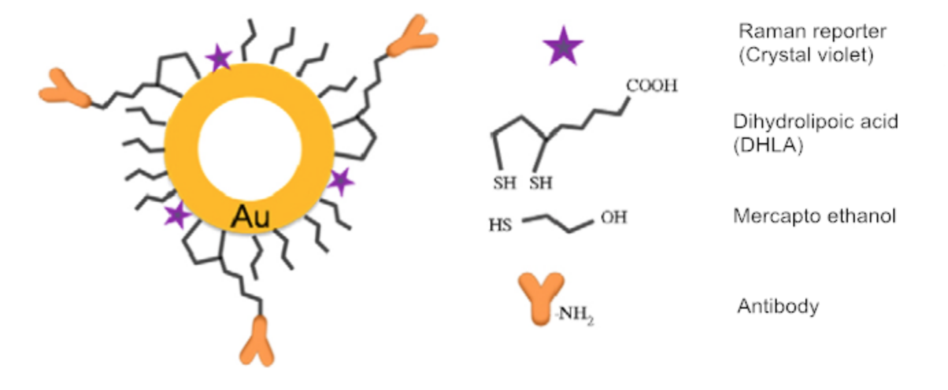 With years of experience in the pharmaceutical and life science sector, Creative Diagnostics launches NanoHollows, which contain cavity enclosed by a non-porous nanoshell. NanoHollows have great potential applications in biological sensing, biomedical imaging and photothermal therapy due to their unique localized surface plasmon resonance (LSPR) feature...
 The system converts laboratory microscopes into powerful tools for live cell imaging to more effectively perform in-depth analysis of cellular mechanisms and behavior in a live environment....
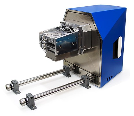 Users of Delmic's SECOM solution for Correlated Light & Electron Microscopy (CLEM) have recently published a review in Nature Methods that illustrates the power of CLEM in the world of biology. The boom in the use of super-resolution microscopies in recent years reached a peak with the award of the 2014 Nobel Prize in Chemistry to Betzig, Hell and Moerner for their contributions in the development of super-resolved fluorescence microscopy...

 École Polytechnique Federale de Lausanne, better known as EPFL, has recently reported on how a group of its scientists have used powerful imaging techniques including nanoIR to support a study which sheds light on photosynthesis. All plants use a form of photosynthesis to produce energy, though not all rely exclusively on it. In higher plants, capturing light takes place in specialized compartments called thylakoids. These are found in cell organelles called chloroplasts, which are the equivalent of a power station for the plant....
 A group of scientists from the CNR is investigating the melting kinetics of poly(3-hydroxybutyrate) (PHB), a natural thermoplastic polymer with mechanical properties comparable to synthetic polymers. PHB is now the focus of scientific interest as it has commercial potential as a fully biodegradable and biocompatible product. Potential uses include environmentally friendly packaging and films, pharmaceutical drug administration, and biocompatible resorbable medical implants. Small batches of PHB have been produced since 1925 by bacterial fermentation, but until now there is no large scale commercial production because it is more expensive than commonly used polymers...
 RJ Lee Group, Inc. is a materials characterization laboratory and industrial forensics consulting firm employing more than 300 scientists, engineers, technicians and support staff at locations in multiple locations across the USA. The firm specializes in scientific support of four areas of interest: analytical laboratory testing, industrial forensics consultation and support, litigation support, and systems reliability tools for engine health....
 The record breaking numbers were drawn by eight parallel conference sessions that embraced the life and physical sciences, and delivered a balanced programme of optical and electron microscopy. This breadth made emc2012 the most inclusive event yet. Professor Tony Wilson, President of the Royal Microscopical Society and Vice Chair of the Congress, said, “The conference sessions have provided an unparalleled opportunity for delegates to immerse themselves in their own area of interest, and also to witness new techniques and tools that might benefit their current work, or feature in their future activities.”...
 The Molecular Engineering Laboratory was established in 2009 by Nobel Laureate Dr Sydney Brenner as a multi-disciplinary laboratory where diverse expertise from different realms of science could come together to develop novel technologies. Post-doctoral fellows in the laboratory undertake a wide variety of research ranging from organic fluorophore development to characterization of marine biomaterials with unique physical properties to genetic therapies.
 Users of the Leica Microsystems Tissue IA product can now source from Indica Labs, a range of image analysis algorithms, providing solutions to many diverse tissue-based quantification applications. Indica Labs' image analysis algorithms for molecular assay and morphological feature detection and quantification in whole slide images, coupled with Leica Microsystems' Tissue IA, provides an easy-to-use solution for complex image analysis problems in Digital Pathology...
 The Institute for Particle Science and Engineering is a centre of excellence within the School of Process, Environmental and Materials Engineering at the University of Leeds. Researchers here focus on projects that address the engineering science of particulate processes and products. PhD and MSc students have been using an LTS350 hot-stage system in their research for several years...
 The Scientific Programme for the 15th European Microscopy Congress includes six plenary talks, 123 invited speakers and 330 oral presentations spread across 72 sessions that embrace light and electron microscopy.
 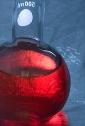 Agar Scientific, a leading supplier of microscopy accessories and consumables, announces a new range of highly concentrated gold colloidal nanoparticles from British Biocell International (BBI). Agar Scientific is a market leader in the supply of high quality accessories to assist with sample handling for the microscopy market serving a very broad range of application areas. One of these ranges is the gold nanoparticles produced by BBI...