
Channels
Special Offers & Promotions
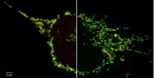
New 2D Superresolution Mode for ZEISS Airyscan Delivers 120 Nanometer Lateral Resolution
Improved optical sectioning delivers higher resolution without the need to acquire a z-stack
At Neuroscience 2017, a new imaging mode for the ZEISS LSM 8 family with Airyscan has been introduced. Their unique 32-channel GaAsP array detector captures more spatial information than traditional confocal microscopes. The new 2D Superresolution mode now uses this additional information to create an optical section of 0.2 Airy units (AU) and resolves structures down to 120 nanometer laterally in a single image.
The benefits for scientists
In the past, researchers had to acquire a stack of z-slices and subsequently deconvolve to get optical sections thinner than one AU and enhance lateral resolution. Temporal resolution was thus limited, and a prolonged light exposure of the sample was inevitable. Scientists can now use the new 2D Superresolution mode to overcome this problem and perform gentle live cell imaging experiments. They profit from very low light exposure, highly resolved structural information and excellent signal-to-noise ratio.
The principle behind
ZEISS Airyscan is an area detector. Unlike traditional confocal microscopes which reject photons from outside of the focal plane at a pinhole, ZEISS Airyscan detects all precious fluorescence emission photons of 1.25 AU. Their information is then used to deliver higher sensitivity, superresolution, and high acquisition speeds. The new 2D Superresolution mode takes advantage of the fact that ZEISS Airyscan captures x, y and z information of the confocal point spread function. A new exclusive processing algorithm uses this inherent spatial information captured in a single image. It specifically distinguishes between photons originating from the focal plane of 0.2 AU and photons from outside of this focal plane. In a traditional confocal microscope, a researcher could only close the pinhole to 0.2 AU to attempt to achieve the same optical sectioning. This would mean sacrificing many photons, even from the focal plane, thus reducing signal-to-noise drastically.
Researchers can process both existing and new ZEISS Airyscan data with the new 2D Superresolution mode.
News Channels
- Latest News
- New Laboratory Products
- Industry News
- Laboratory Automation | IT Solutions
- Microscopy | Image Analysis
- Separation Science
- Research | Case Studies
- Video Presentations
- Events | Conferences
Subscribe to any of our newsletters for the latest on new laboratory products, industry news, case studies and much more!

Popular this Month
Top 10 most popular articles this month
Today's Picks
Looking for a Supplier?
Search by company or by product
Please note Lab Bulletin does not sell, supply any of the products featured on this website. If you have an enquiry, please use the contact form below the article or company profile and we will send your request to the supplier so that they can contact you directly.
Lab Bulletin is published by newleaf marketing communications ltd.
Media Partners