
Channels
Special Offers & Promotions
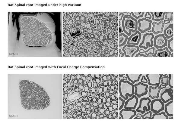
New Focal Charge Compensation mode for ZEISS field emission scanning electron microscopes improves image quality
 In collaboration with the National Center for Microscopy and Imaging Research (NCMIR) at the University of California San Diego, ZEISS releases a new Focal Charge Compensation module for block face imaging with ZEISS GeminiSEM and 3View® from Gatan, Inc.
Focal Charge Compensation expands versatility and considerably increases data quality without prolonging acquisition times and enables easy imaging of even the most charge-prone samples. Resin-embedded tissues and cells can be imaged without charging artifacts, while the pixel dwell time is reduced. Decreasing beam exposure time not only ensures fast acquisition rates, but also guards against sample damage, which is key to acquiring reliable and reproducible 3D data. Professor Mark H. Ellisman, Director of NCMIR, says “Focal Charge Compensation will breathe new life into block face scanning electron microscopy by allowing high quality imaging of previously intractable specimens, including legacy samples prepared with traditional electron microscopy stains.”
Preventing charging effects
This extension of the 3View® system from Gatan, Inc eliminates specimen charging. A gas injection system consisting of a tiny capillary needle is precisely located above the sample. Nitrogen is guided through this needle directly onto the block face surface while the chamber is maintained under high vacuum. This eliminates charging without degrading image quality. The needle retracts automatically during the cutting cycle so the workflow is uninterrupted and high acquisition rates are maintained.
Producing thousands of serial images in a single day
The 3View® system consists of an ultramicrotome directly integrated into the vacuum chamber of the ZEISS Sigma and ZEISS GeminiSEM field emission scanning electron microscopes. It enables automated serial block face imaging of embedded samples (e.g. cells or tissue) with a slice thickness down to 15 nanometers. The sample is continuously cut and imaged, and a three-dimensional rendering of the sample with nanometer-scale resolution can be reconstructed.
News Channels
- Latest News
- New Laboratory Products
- Industry News
- Laboratory Automation | IT Solutions
- Microscopy | Image Analysis
- Separation Science
- Research | Case Studies
- Video Presentations
- Events | Conferences
Subscribe to any of our newsletters for the latest on new laboratory products, industry news, case studies and much more!

Popular this Month
Top 10 most popular articles this month
Today's Picks
Looking for a Supplier?
Search by company or by product
Please note Lab Bulletin does not sell, supply any of the products featured on this website. If you have an enquiry, please use the contact form below the article or company profile and we will send your request to the supplier so that they can contact you directly.
Lab Bulletin is published by newleaf marketing communications ltd.
Media Partners