
Channels
Special Offers & Promotions
PI Positioning Stages Help Reveal Secrets of Teeth
Tooth decay is often revealed by an X-ray taken in the dentist’s chair.
 The image taken is relatively coarse and only reveals the presence of the problem, what it doesn’t show is detail about the mechanism that has led to the decay. Understanding the processes that occur during tooth decay and indeed repair involves researchers looking at concentrations of minerals such as Calcium present in the structure. X ray microtomography is now helping researchers at Queen Mary, University of London (QMUL) gain a deeper understanding of these underlying processes by generating high resolution images that can detail mineral concentrations in the tooth. The image generation involves scanning the tooth and X-ray detector with high precision over an extended time period. PI linear and rotary stages, chosen for their precision and long term reliability, have been successfully employed in this research work for a number of years.
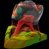
X-ray microtomography uses X-rays to build up a virtual 3D model of an object from a series of two dimensional scans. In the research, headed up by Dr Graham Davis, School of Dentistry at QMUL, microtomography is combined with the long X-ray exposure of extracted teeth to enhance as much as possible the contrast ratio of the image. The resulting model reveals details of both the structure and density of the tooth. The grey levels of the image are calibrated to identify the density at any given point and this in turn with priori information about the composition can be used to show mineral concentrations to a precision of better than 1%. By observing the tooth over an extended time period, changes in mineral concentration resulting from further decay or re-mineralisation treatment can be monitored. This in turn helps a researcher understand how treatments work, what treatments are effective and how specific treatments can be managed.
The longer the exposure time of a scan, the higher the contrast ratio, the better the mineral concentration that can be measured. With scans taking several hours the mechanical stages used need to be reliable over the long term.
Three PI M-500 series high precision linear stages with recirculating ball guides and a PRS-110 high precision rotary stage with exceptionally low wobble are used in combination to scan the sample and X ray detector. One of the linear stages is used to scan the lead encased detector while the other two linear stages move the sample in the Z and focal planes. The rotary stage is used to index the sample after each scan. The detector moves dynamically with image collection triggered by position information from the linear stage. The exceptional velocity stability, repeatability, straightness and flatness of the mechanics are the key to QMUL being able to generate the high resolution images that are subsequently combined into the computer generated 3D model of the tooth.
The work at QMUL has been ongoing for nearly 20 years with the first stages provided by PI still in operation. Results have shown that by observing mineral concentrations of less than 1%, details about how a tooth decays or repairs under treatment can be better observed and understood.
PI would like to thank Dr Graham Davis and David Mills of The School of Dentistry at Queen Mary, University of London for their help in writing this article.
News Channels
- Latest News
- New Laboratory Products
- Industry News
- Laboratory Automation | IT Solutions
- Microscopy | Image Analysis
- Separation Science
- Research | Case Studies
- Video Presentations
- Events | Conferences
Subscribe to any of our newsletters for the latest on new laboratory products, industry news, case studies and much more!

Popular this Month
Top 10 most popular articles this month
Today's Picks
Looking for a Supplier?
Search by company or by product
Please note Lab Bulletin does not sell, supply any of the products featured on this website. If you have an enquiry, please use the contact form below the article or company profile and we will send your request to the supplier so that they can contact you directly.
Lab Bulletin is published by newleaf marketing communications ltd.
Media Partners