
Channels
Special Offers & Promotions
Tomocube
Products
Contact Tomocube
All articles from Tomocube
CrestOptics and Tomocube partner to advance 3D imaging with new multimodal imaging platform
Nov 20, 2024
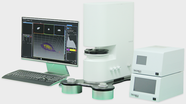
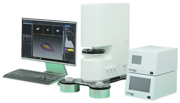
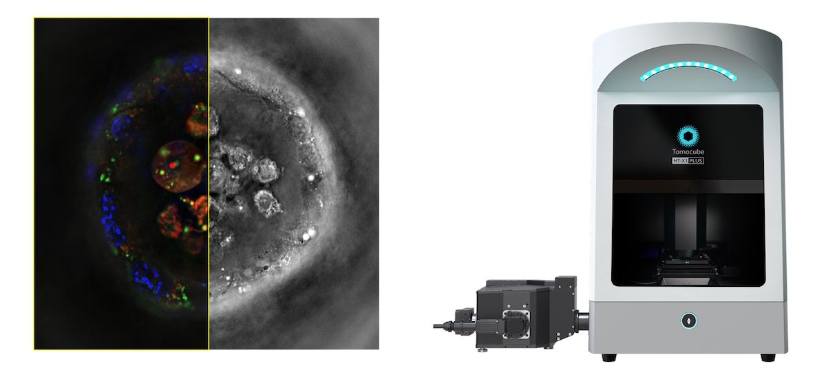 CrestOptics S.p.A., a manufacturer of high-end microscopy solutions and advanced systems for fluorescence microscopy, and Tomocube, a leader in holotomography (HT) technology, today announced a strategic collaboration to provide a next-generation multimodal imaging platform, the HT-X1™ Plus. By combining the expertise of both companies, this platform integrates CrestOptics’ spinning disk confocal technology with Tomocube’s latest innovation...
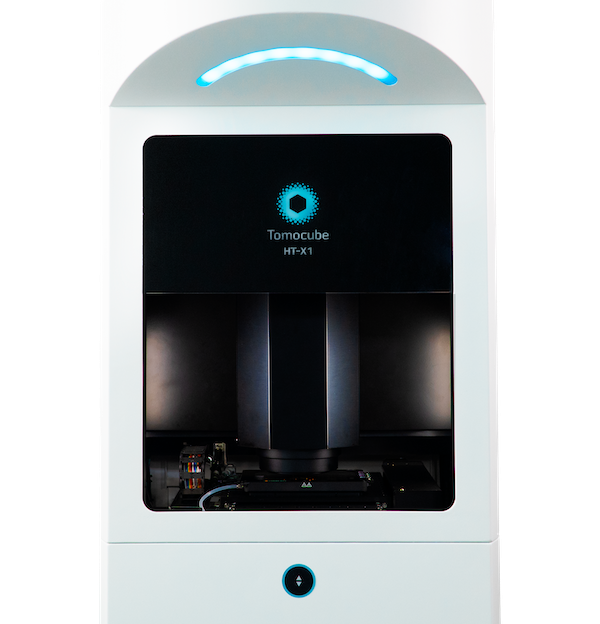 Ground-breaking technology unlocks label-free 3D and 4D live cell imaging on standard imaging plates for higher-throughput and automated screening applications. A novel optical microscope utilizing incoherent light to generate holographic images of unlabelled live cells is now available from Tomocube. Called HT-X1, the new microscope is ideally suited to higher-throughput and automated screening applications with its ability to image multi-well plate formats, large field-of-view, laser autofocus system, and very high performance 0.95NA objective...
 Tomocube’s cutting-edge 3D quantitative phase imaging is set to play a crucial role in platelet research according to a new paper from scientists at the University of York. Using holotomography images generated by the Tomocube HT-2H microscope, the team were able to identify and quantify in single unlabelled, live platelets clear disparities in activation status and potential functional ability in disease states without experimental interference, such as from fixation or labelling...
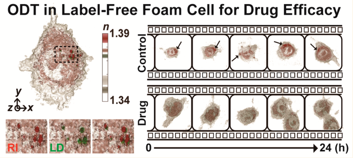 Label-free holotomography enables quantitative morphological and biophysical analysis of Atherosclerosis foam cells and precise measurement of targeted nanodrug effectiveness at single-cell level. Treatment of Atherosclerosis, the disease responsible for approximately half of all deaths in westernised societies, is a step closer following correlative studies using Tomocube’s HT-2 microscope....
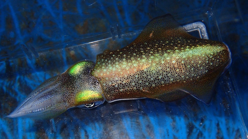 The seemingly effortless and instantaneous colour and iridescence changes displayed by octopuses, squid and cuttlefish is one of Nature’s most amazing sights. Now, researchers have discovered how to use the cephalopod proteins responsible for these changes to manipulate the optical refractive index (RI) of mammalian cellular compartments. By changing this intrinsic physical property of the cell, microscopists could soon be using the RIs of live cells as molecular probes...
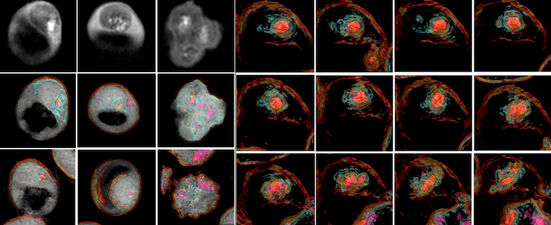 3D label-free imaging identifies real-time cholesterol sorting mechanism in Plasmodium falciparum-infected human erythrocytes. The first study to elucidate the sequential dynamics of membrane cholesterol transport in erythrocytes infected with live Plasmodium falciparum parasites has been successfully concluded using the 3D label-free imaging capability of holotomography microscopy....
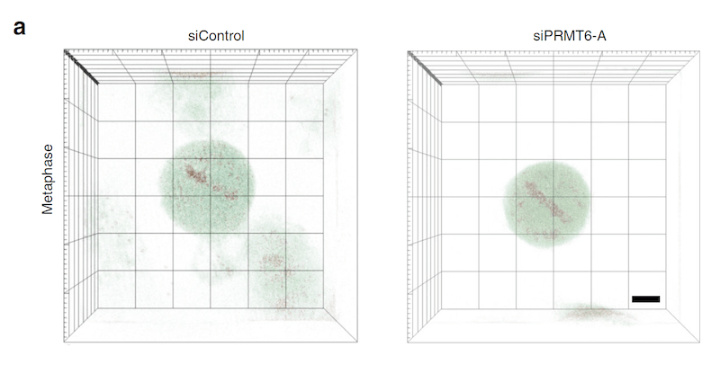 Governing molecular mechanism for chromosome condensation identified 140 years after mitosis first described. The molecular mechanism underlying chromosome condensation, one of the key steps in cell division, has been discovered from three-dimensional (3-D) refractive index (RI) reconstructions of individual mitotic cells. Writing in Nature Communications1, the authors describe using the Tomocube HT-1S holotomographic microscope to quantify the structural and biochemical parameters of the cytoplasm and chromosomes within the individual mitotic cells...
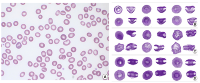 Uniquely shaped red blood cells observed for the first time in patient blood could be breakthrough in diagnosis of Myelodysplastic Syndrome (MDS). Although the three-dimensional (3-D) shape of erythrocytes is strongly associated with various malignant blood disorders, conventional optical imaging approaches only provide information on two-dimensional morphology. Now, a Korean research team has used the Tomocube holotomography microscope to observe uniquely shaped red blood cells (RBCs) for the first time....
Tomocube Holotomography Microscopy enables Label-free, Non-invasive Study of individual Spermatozoon
Dec 3, 2019
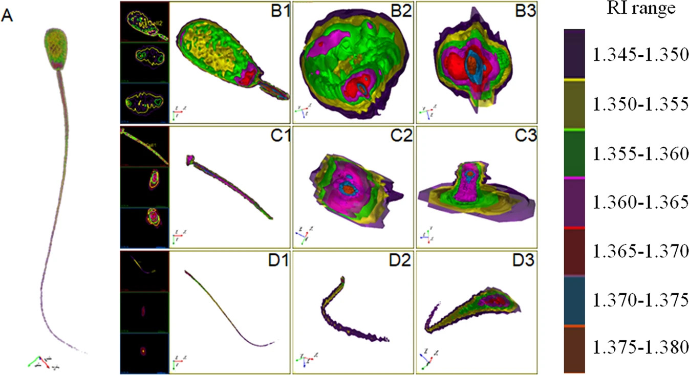 Rapid 3D imaging and in-depth structural analysis opens up quality monitoring and spermatozoa selection for artificial insemination in humans and animals. A key determinant for success in artificial insemination for humans and animals is the quality of the spermatozoa. However, current 2D label-free imaging methods can only detect abnormal spermatozoa with significant morphological changes or incomplete structures and are unable to provide information on their internal structure or composition....
 Development of the world’s first holotomographic microscope with 3D fluorescence recognised as photonics breakthrough by the 2019 Microscopy Today Innovation Award. Disruptive technology that combines the quantitative phase imaging (QPI) approach of label-free 3D refractive index (RI) tomography with fluorescence imaging to reveal the structure, volume, surface area, concentration, and dry matter mass of individual live cells in real-time....
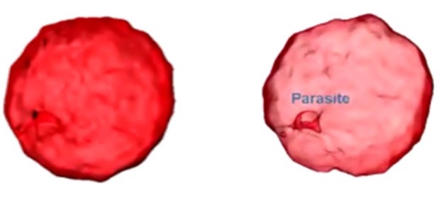 Recently, a number of studies have been published demonstrating that label-free, 3-D imaging using holotomography (HT) microscopy enables researchers to observe morphological and chemical alterations of host cells due to the parasite infection without any transfection or dye staining. This powerful new tool allows parasites to be easily and quickly detected and monitored within the host cells and permits the intricacies of parasite infection mechanisms and the host cell/parasite life cycle to be studied....
Tomocube 3D Microscope Delivers Breakthrough in Label-Free Quantification of Intracellular Lipids
Jul 18, 2019
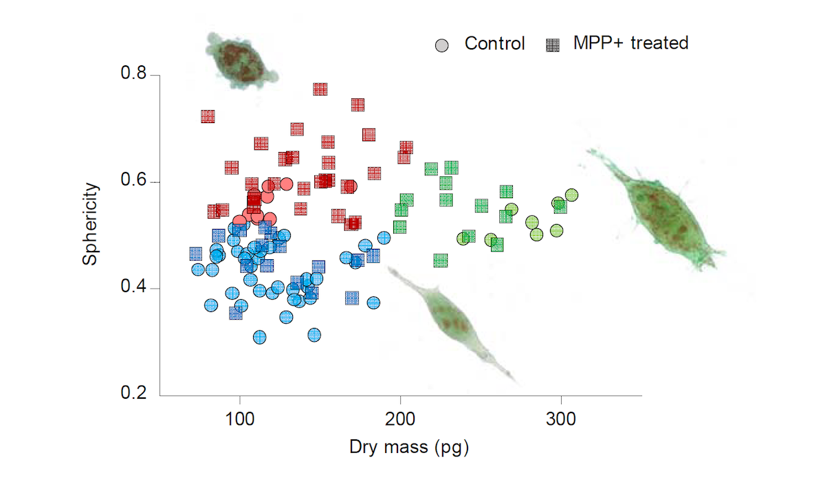
Tomocube 3D Microscope Captures Key Cellular Dynamic Responses to Parkinson Drug Treatment
Mar 20, 2019
 Motorized stage and custom-designed stitching software implemented across the range deliver large 4D images to advance long term live cell imaging studies. Long-term studies of live cells are set to take a significant leap forward with the introduction of a motorized stage and custom-designed stitching software for Tomocube’s holotomography microscopes. The automated combination can be fitted to every microscope model in the company’s range and produces large field-of-view images and time-lapse videos....
 Global support for Holotomography Microscopy as company prepares for introduction of second microscope model. A comprehensive network of specialist imaging companies has been unveiled to support the sales and maintenance of Tomocube’s holotomography microscopes around the world. Covering North America, Europe and Asia, the 23-strong group is being launched as Tomocube prepares to introduce its second microscope model in less than three months....
 Tomocube’s revolutionary holotomography technique delivers 3D structural and chemical information quickly and simply without any labelling or pre-preparation. A spin-out company from the renowned Korea Advanced Institute of Science and Technology in Daejeon has been established to commercialize the revolutionary Holotomography (HT) microscopy developed...
Media Partners