LaVison BioTec introduce new products for their UltraMicroscope and TriM Scope microscopy products at the 2014 annual meeting of the Society for Neuroscience.
LaVision BioTec announce the release of several ground-breaking new products at the 2014 meeting of the Society for Neuroscience to be held in Washington DC, November 15-19. Delegates are invited to visit LaVision on Booth #817 to meet the product group and learn about the latest innovations from the company. TriM Scope II is an intravital 2-photon microscope. Three new developments are announced at the meeting. First is a...
With the new Olympus cellSens imaging software offering new functions and support for the latest microscopy hardware, life science researchers can now observe and react to the most elusive events taking place within biological samples.
Assisting researchers in gaining a deeper understanding of dynamic biological processes, the new cellSens imaging software (version 1.12) ensures the most efficient use of valuable time-lapse experiments and the latest microscopy hardware. Building on the capabilities introduced by Olympus with its unique Graphical Experiment Manager (GEM) interface, cellSens 1.12 allows the user to truly get in touch with their sample. Enabling effortless...
Market leaders in temperature controlled microscopy, Linkam Scientific Instruments, are working with French instrument company, Xenocs, to provide the facility to perform X-ray experiments under changing temperature and/or tensile conditions.
Linkam is pleased to announce that their temperature stages have been chosen by Xenocs to incorporate in their recently launched Nano-inXider X-ray characterisation system opening new perspectives in many research fields. Xenocs is a French instrument company formed as a spinout from the Laue Langevin Institute. The company provides solutions for nanomaterial characterisation using Small and Wide Angle X-ray Scattering technique...
A novel super-resolution fluorescence microscope equipped with a low-noise, high-speed 5.5 Megapixel Andor Neo sCMOS camera has enabled the real-time nanoscopic imaging of large fields of living cells for the first time.
Researchers from the Max Planck Institute in Goettingen, Germany, adopted massive parallelisation techniques to create 116,000 simultaneous scanning points and super-resolve 120 µm × 100 µm fields in less than a second. The research was led by Professor Stefan Hell, who first advanced STED (Stimulated Emission Depletion) and RESOLFT (Reversible Saturable/Switchable Optical Fluorescence Transitions) far-field, super-resolution microscopy...
JPK Instruments reports on the use of the JPK NanoWizard® AFM system at ITAV, the Institut des Technologies Avancées en science du Vivant, in Toulouse in the South of France.
Dr Childérick Severac manages the Bionanotechnologies Platform at ITAV (Institut des Technologies Avancées en science du Vivant) on the site of the new Oncopole campus dedicated to cancer research. His research activities may be divided into two parts: his own research and the research activity he does for others in the form of providing a collaborative imaging service. Dr Severac's research focuses on the development of...
Highest speed for brain research with ZEISS MultiSEM 5
At the annual Neuroscience meeting in Washington, D.C., November 15-19, 2014, ZEISS will present ZEISS MultiSEM 505, its new scanning electron microscope (SEM). As the first SEM in the world, the system features 61 beams working in parallel, and offers an unrivaled capture speed of 1220 megapixels per second at a pixel size of 4 nm. This high acquisition speed is used for imaging neural tissue in brain research where it is now possible to observe much bigger...
Syngene is delighted to introduce the vibrant new T:Genius imaging system, which unlike any other imager on the market, allows high performance, walk-away imaging of gels and blots on a smartphone or tablet.
What makes the new T:Genius outperform other commercial imagers is the amazing ability to quickly and easily access stunning quality images anywhere from a tablet, computer or smartphone. Using the T:Genius’ clever ‘StatusLink’ feature researchers can stay updated and even share image results with colleagues in other labs, no matter whether they are down the hall or in another country. This is a great time saver when, for instance, scientists are...
Building on the success and principles of SMART Automation, Sakura Finetek proudly introduces the next step in Total Laboratory Automation: Tissue-Tek® AutoSection® Automated Microtome, advancing histopathology laboratories and improving patient care.
PIE Scientific, a leader in low pressure high efficiency plasma source manufacturing, has recently released a revolutionary downstream plasma cleaner---SEMI-KLEEN plasma cleaner.
It is designed to meet the toughest contamination requirements from the semiconductor capital equipment industry. The revolutionary low plasma potential discharge technique greatly reduces the risk of ion sputtering damage. Together with a proprietary two-stage filter gas delivery system, SEMI-KLEEN plasma cleaner has been proven capable of meeting particle contamination requirement for sub 10nm nodes....
High Throughput Screening for Raman chemical analyzers
HORIBA Scientific, a leading innovator in the manufacture of high performance scientific and analytical equipment, is announcing the release of its new MultiWell module for the acclaimed LabSpec 6 Spectroscopy Suite. The module transforms the HORIBA portfolio of Raman microscopes into efficient high throughput chemical analyzers, coupling the non-destructive chemically rich analysis with high sample turn around and fast return on...
JPK Instruments reports on the use of their AFM system, the NanoWizard®, in the Nanobiosciences Group of the Institute of Physical & Theoretical Chemistry at the Technical University of Braunschweig.
It is being used to study DNA and DNA nanostructures. Dr Philip Tinnefeld is Professor of Biophysical Chemistry at the Technische Universität Braunschweig where he runs a laboratory which specialises in the field of NanoBioScience. His group has pioneered the integration of DNA origami with optical single-molecule spectroscopy over the last few years. In this context, they have demonstrated some of the early applications where the breakthroughs of...
Improved imaging tools to better understand the brain
The National Institutes of Health (NIH) announced its first research grants through President Barack Obama's BRAIN Initiative, including three awards to the University of California, Berkeley, totaling nearly $7.5 million over three years. In addition, a $12 million public-private collaboration between ZEISS and UC Berkeley to support the Berkeley Brain Microscopy Innovation Center (BrainMIC) was made public. The US Brain initiative is focused on advancing tool development in...
ZEISS congratulates Eric Betzig, Stefan Hell and William Moerner on winning the 2014 Nobel Prize for Chemistry.
All three underscored the value of microscopy in research with their discovery of methods to achieve superresolution. The limit of optical resolution calculated by Ernst Abbe, company founder Carl Zeiss's partner, in the 19th century has been considerably improved through the use of modern technologies. Today, photo-activated localization microscopy (PALM) enables scientists to view processes in living cells and tissues down to the molecular level – in other words, with resolution in...
Cytation™ 5 is a uniquely integrated, configurable system that combines automated digital widefield microscopy with conventional multi-mode microplate detection to provide phenotypic cellular information and well-based quantitative data.
LaVison BioTec, developers of advanced microscopy solutions for the life sciences, report on users of their Ultramicroscope Light Sheet Fluorescence Microscope system to aid the research of the Miami Project to Cure Paralysis under the supervision of Professor Vance Lemmon, the Walter G. Ross Distinguished Chair in Developmental Neuroscience & Professor of Neurological Surgery at the University of Miami.
In 2003, Professor Vance Lemmon accepted a position at The Miami Project to Cure Paralysis at the University of Miami. This centre has taken the philosophy that by promoting interactions between basic and clinical scientists, it will be possible to speed the finding of a cure for a devastating clinical problem. This research has focused on answering questions that help define human spinal cord injury and reveal strategies for the repair of damaged spinal tissue...
Introducing the 5 megapixel DP27 and 3 megapixel DP22, Olympus presents its next generation of digital cameras for microscopy. Individual needs are met with multi-mode functionality, delivering Full-HD live images of unprecedented quality directly to the monitor.
Life-like digital microscopy is now a reality with the DP27 and DP22 digital cameras from Olympus. Both cameras deliver fluid imaging at Full-HD resolution via a USB 3.0 interface, and enable easy optimisation for each and every application with three distinct modes. With the next generation of Olympus digital microscopy camera, researchers can enjoy visualisation on a monitor that is virtually identical to that of the oculars....
Meet world’s first Mobile SEM - the Phenom desktop SEM. A fully integrated and accredited scanning electron microscope (SEM) with EDX to be used on every location for fast and accurate investigations and analysis.
The Phenom ProX is the ultimate all-in-one imaging and X-ray analysis system (EDX). With the Phenom ProX desktop SEM, sample structures can be physically examined and their elemental composition determined. Besides point analysis, the optional Elemental Mapping and Line Scan software allows further analysis of the distribution of elements....
Use of Picsara will provide a more streamlined and secure approach to the patient journey.
HCA International, providers of world class private hospital care, are to collaborate with Swedish-based medical imaging specialists Mawell to transform the delivery of frontline care, enhance their service to patients and improve clinical safety and security. Launching across all HCA International’s clinical UK sites early next year Picsara, a unique multidisciplinary imaging software solution, will enable medical staff across various specialities to capture, store, distribute, analyse, annotate and measure all non-radiology medical images and videos efficiently and securely to help...
Thanks to a novel software upgrade from Olympus, an optical resolution of up to 120 nm is now achieved in cell and tissue imaging.
With the new Olympus FV-OSR software, the FluoView FV1200 is transformed into a powerful system for confocal Super Resolution microscopy. Introducing a smart route to Super Resolution microscopy, the Olympus FV-OSR software module enables users of the FluoView FV1200 easy access to what was previously the domain of only specialised microscopy systems. The extra detail offered by Super Resolution technology facilitates researchers in...
Xmark Media the organisers of Photonex 2014, the UK’s showcase photonics conference & exhibition announce the programme and speakers for the meeting on Nano and Bio-Imaging.
This FREE-to-attend conference will be held on Wednesday 15th October as part of a diverse two-day event at the visitor-friendly Ricoh Arena in Coventry. Applying photonics to a broad group of imaging techniques is providing greatly increased information particularly in the fields of medicine and the life sciences. With researchers continually looking for increased resolution with the addition of chemical information, this year’s conference provides a comprehensive overview of this rapidly expanding field....
Syngene, a world-leading manufacturer of image analysis solutions, today announced its G:BOX Chemi XT4 is being used by scientists at the University of Edinburgh to accurately quantify DNA and proteins from bacteria engineered to produce metal nanoparticles.
This work is contributing to speeding up throughput of research on novel methods of processing toxic metals from contaminated soil. Researchers in the Horsfall Laboratory at the University of Edinburgh in Scotland are using a G:BOX Chemi XT4 multi-application imager to analyse agarose gels of bacterial DNA stained with the green fluorescent dye SafeView™. They are also utilising the G:BOX Chemi XT4 to image SDS-PAGE gels and...
The Royal Swedish Academy of Sciences has decided to award the Nobel Prize in Chemistry for 2014 to Eric Betzig, Janelia Farm Research Campus, Howard Hughes Medical Institute, Stefan W. Hell, Max Planck Institute for Biophysical Chemistry and German Cancer Research Center and William E. Moerner, Stanford University, Stanford, CA, USA
“for the development of super-resolved fluorescence microscopy”.


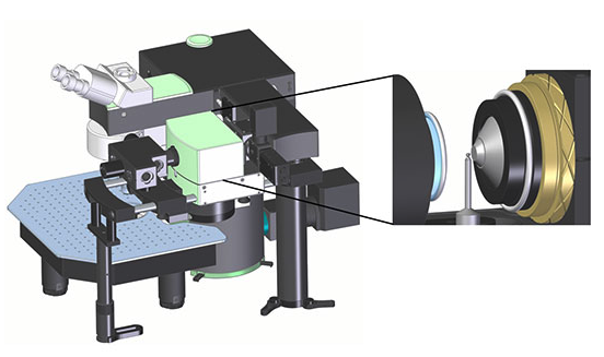 LaVision BioTec announce the release of several ground-breaking new products at the 2014 meeting of the Society for Neuroscience to be held in Washington DC, November 15-19. Delegates are invited to visit LaVision on Booth #817 to meet the product group and learn about the latest innovations from the company. TriM Scope II is an intravital 2-photon microscope. Three new developments are announced at the meeting. First is a...
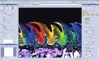 Assisting researchers in gaining a deeper understanding of dynamic biological processes, the new cellSens imaging software (version 1.12) ensures the most efficient use of valuable time-lapse experiments and the latest microscopy hardware. Building on the capabilities introduced by Olympus with its unique Graphical Experiment Manager (GEM) interface, cellSens 1.12 allows the user to truly get in touch with their sample. Enabling effortless...
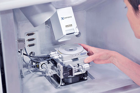 Linkam is pleased to announce that their temperature stages have been chosen by Xenocs to incorporate in their recently launched Nano-inXider X-ray characterisation system opening new perspectives in many research fields. Xenocs is a French instrument company formed as a spinout from the Laue Langevin Institute. The company provides solutions for nanomaterial characterisation using Small and Wide Angle X-ray Scattering technique...
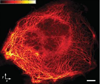 Researchers from the Max Planck Institute in Goettingen, Germany, adopted massive parallelisation techniques to create 116,000 simultaneous scanning points and super-resolve 120 µm × 100 µm fields in less than a second. The research was led by Professor Stefan Hell, who first advanced STED (Stimulated Emission Depletion) and RESOLFT (Reversible Saturable/Switchable Optical Fluorescence Transitions) far-field, super-resolution microscopy...
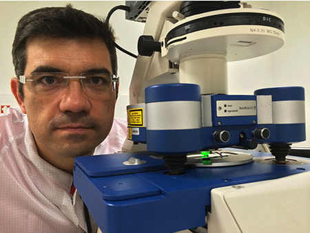 Dr Childérick Severac manages the Bionanotechnologies Platform at ITAV (Institut des Technologies Avancées en science du Vivant) on the site of the new Oncopole campus dedicated to cancer research. His research activities may be divided into two parts: his own research and the research activity he does for others in the form of providing a collaborative imaging service. Dr Severac's research focuses on the development of...
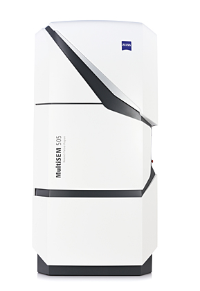 At the annual Neuroscience meeting in Washington, D.C., November 15-19, 2014, ZEISS will present ZEISS MultiSEM 505, its new scanning electron microscope (SEM). As the first SEM in the world, the system features 61 beams working in parallel, and offers an unrivaled capture speed of 1220 megapixels per second at a pixel size of 4 nm. This high acquisition speed is used for imaging neural tissue in brain research where it is now possible to observe much bigger...
 What makes the new T:Genius outperform other commercial imagers is the amazing ability to quickly and easily access stunning quality images anywhere from a tablet, computer or smartphone. Using the T:Genius’ clever ‘StatusLink’ feature researchers can stay updated and even share image results with colleagues in other labs, no matter whether they are down the hall or in another country. This is a great time saver when, for instance, scientists are...
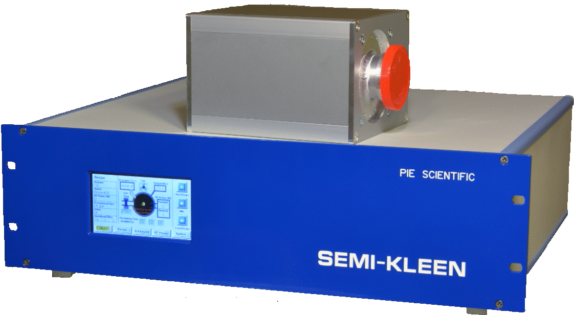 It is designed to meet the toughest contamination requirements from the semiconductor capital equipment industry. The revolutionary low plasma potential discharge technique greatly reduces the risk of ion sputtering damage. Together with a proprietary two-stage filter gas delivery system, SEMI-KLEEN plasma cleaner has been proven capable of meeting particle contamination requirement for sub 10nm nodes....
 HORIBA Scientific, a leading innovator in the manufacture of high performance scientific and analytical equipment, is announcing the release of its new MultiWell module for the acclaimed LabSpec 6 Spectroscopy Suite. The module transforms the HORIBA portfolio of Raman microscopes into efficient high throughput chemical analyzers, coupling the non-destructive chemically rich analysis with high sample turn around and fast return on...
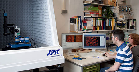 It is being used to study DNA and DNA nanostructures. Dr Philip Tinnefeld is Professor of Biophysical Chemistry at the Technische Universität Braunschweig where he runs a laboratory which specialises in the field of NanoBioScience. His group has pioneered the integration of DNA origami with optical single-molecule spectroscopy over the last few years. In this context, they have demonstrated some of the early applications where the breakthroughs of...
 The National Institutes of Health (NIH) announced its first research grants through President Barack Obama's BRAIN Initiative, including three awards to the University of California, Berkeley, totaling nearly $7.5 million over three years. In addition, a $12 million public-private collaboration between ZEISS and UC Berkeley to support the Berkeley Brain Microscopy Innovation Center (BrainMIC) was made public. The US Brain initiative is focused on advancing tool development in...
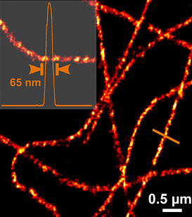 All three underscored the value of microscopy in research with their discovery of methods to achieve superresolution. The limit of optical resolution calculated by Ernst Abbe, company founder Carl Zeiss's partner, in the 19th century has been considerably improved through the use of modern technologies. Today, photo-activated localization microscopy (PALM) enables scientists to view processes in living cells and tissues down to the molecular level – in other words, with resolution in...
 In 2003, Professor Vance Lemmon accepted a position at The Miami Project to Cure Paralysis at the University of Miami. This centre has taken the philosophy that by promoting interactions between basic and clinical scientists, it will be possible to speed the finding of a cure for a devastating clinical problem. This research has focused on answering questions that help define human spinal cord injury and reveal strategies for the repair of damaged spinal tissue...
.")
 With the new Olympus FV-OSR software, the FluoView FV1200 is transformed into a powerful system for confocal Super Resolution microscopy. Introducing a smart route to Super Resolution microscopy, the Olympus FV-OSR software module enables users of the FluoView FV1200 easy access to what was previously the domain of only specialised microscopy systems. The extra detail offered by Super Resolution technology facilitates researchers in...
 This work is contributing to speeding up throughput of research on novel methods of processing toxic metals from contaminated soil. Researchers in the Horsfall Laboratory at the University of Edinburgh in Scotland are using a G:BOX Chemi XT4 multi-application imager to analyse agarose gels of bacterial DNA stained with the green fluorescent dye SafeView™. They are also utilising the G:BOX Chemi XT4 to image SDS-PAGE gels and...