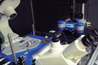
Trio of Andor iXon EMCCD cameras on Revolution XD microscope make possible the simultaneous tracking of key autophagosome markers to deliver new insights into organelle biogenesis
The location of the site where autophagosomal membranes are created within the cell has been the subject of two competing models for many years. Now, a group of researchers from Osaka University led by Prof. Maho Hamasaki has located the origin of autophagosomes within the cell and shown that neither model is correct. Writing in Nature, they show that autophagosomes form at the contact site of the endoplasmic reticulum (ER) with mitochondria. Live cell imaging studies, using an Andor Revolution XD spinning disk confocal microscope equipped with three Andor iXon 3 EMCCD cameras, captured previously hidden detail...
TissueMark software automatically marks and detects potentially cancerous sections in tissue samples with unprecedented levels of speed and accuracy.
Leap in precision equates to the shift from a freehand outline sketch of a country’s border to a high resolution map of the whole country. New technology, developed in Belfast, will soon enable pathologists to automate the process of marking tissue samples with unprecedented accuracy. TissueMark, developed by Belfast-based digital pathology specialists PathXL, is being launched today at the Association for Molecular Pathology conference in Phoenix, Arizona. TissueMark analyses the detailed structural patterns in tissue samples and marks the boundaries of potentially cancerous sections for more detailed analysis. The new software will help accelerate cancer research and discovery, reduce time in drug development and identify new markers of the disease...
Phenom World has launched a new software – ParticleMetric, for the visualization and analysis of particles and powders.
The combination of ease of use and superb imaging quality of the Phenom desktop SEM with the imaging and particle analysis of ParticleMetric creates a powerful tool for inspecting a wide range of particle and powder samples. The Phenom desktop SEM with ParticleMetric software allows easy generation and analysis of SEM images. The integrated ParticleMetric software allows the user to gather morphology and particle size data for many submicron particle applications. The fully automated measurements of ParticleMetric allow a level of visual exploration beyond optical microscopy that can lead to new discoveries and innovations in powder design, development, and quality control.....
Sensitive, flexible, field-upgradeable system supports a wide range of applications
Molecular Devices®, the market leader in microplate reader technology for over 25 years, has announced today the next-generation SpectraMax® MiniMax™ 300 Imaging Cytometer. This MiniMax 300 imaging cytometer enables both cellular visualization and first-of- its-kind stain free cell-based analysis on the field-upgradable SpectraMax® i3 Multi-Mode Microplate Reader. Brightfield and fluorescence based green and red channel cellular image acquisition and analysis is made simple using the SoftMax Pro® software workflow....
Specialised Imaging reports on how its SIM-D8 ultra high-speed framing camera is being used by Sorlox Corporation (Irvine, CA, USA) to look at the behaviour of plasma generated by a compact Magnetised Target Fusion (MTF) system.
Sorlox Corporation is a pioneer in the development of compact pulsed-plasma devices for use as highly efficient, environmentally friendly power production or for medical isotope production. Using Deuterium as its fuel source - the Sorlox Nautilus Compressor employs an electric field to generate a hot plasma. Using a magnetic field, the Nautilus system compresses the plasma to a high energy state, facilitating fusion and releasing heat that can be used for power generation...
Market leaders in temperature controlled microscopy, Linkam Scientific Instruments report on the use of their innovative CMS196 cryo stage for the study of mammalian cells at the London Research Institute, Cancer Research UK.
For mammalian cells to remain in a healthy state, they require constant renewal of their components. The process of disposing of old components is known as 'autophagy', which stems from the Greek words auto "self" and phagein "eat". This process involves the formation of a double-membrane structure called an autophagosome, which engulfs old or dysfunctional organelles and then fuses with lysosomes, where they are broken down to recycle the constituent molecules....
Live Cell CCD Camera Alternative Captures Frame Rates 10 Times Faster
optiMOS is suited for a broad range of fluorescence microscopy applications. The camera is ideal for cell biologists using live cell, multicolor fluorescence; biophysicists studying membrane dynamics and protein and lipid trafficking; as well as neuroscientists looking at ion transport such as electrophysiology, calcium imaging and ratiometric imaging.
Read MoreLimited Hospital Budgets Prepare Europe for a Breakthrough in Refurbished Medical Imaging EquipmentDec 5, 2013
Already popular in private healthcare, increasing global acceptance of refurbished equipment encourages uptake in the public sector.
The Eurozone economic slowdown has made it tough for hospitals, particularly in Southern Europe, to procure new imaging modalities. With streamlined budgets As a result, refurbished systems are now preferred across most imaging departments, including nuclear medicine, mammography and minimally invasive surgeries in private hospitals. New analysis from Frost & Sullivan’s Analysis of the European Refurbished Medical Imaging Equipment Market finds the market earned revenue of $417.6 million “Growth of the private healthcare sector has increased the uptake of high-quality imaging modalities, as private hospitals focus strongly on returns on investment,” said Frost & Sullivan Healthcare...
Read MoreYeast Species Identification from Positive Blood Culture in just 20 minutesDec 5, 2013
New Candida QuickFISH BC Rapid Molecular Diagnostic Test Now Available
The new Candida QuickFISH BC Rapid Molecular Diagnostic Test can positively identify C. albicans, C. glabrata, and C. parapsilosis, directly from yeast-positive blood cultures in just 20 minutes. The CE marked, nucleic acid based test enables the microbiologist to report pathogen-identification results 2 to 5 days earlier than conventional methods. This enables clinicians, consultant microbiologists and hospital pharmacists to better optimise therapy for patients with fungaemia....
New Technology Could Be The Greatest Breakthrough In Cancer Treatment
TissueMark software automatically marks and detects potentially cancerous sections in tissue samples with unprecedented levels of speed and accuracy. Technology has the potential to save pathologists a combined 250,000 working days of effort per annum. Leap in precision equates to the shift from a freehand outline sketch of a country’s border to a high resolution map of the whole country. New technology, developed in Belfast, will soon enable pathologists to automate the process of marking tissue samples with unprecedented accuracy. TissueMark, developed by digital pathology specialists PathXL, analyses the detailed structural patterns in tissue samples and marks the boundaries of potentially cancerous sections for more detailed analysis...
Read MoreSciGene Adds Sodium Thiocyanate Pretreatment Reagent to FISH Product LineDec 4, 2013
SciGene today introduced its new Sodium Thiocyanate Pretreatment Reagent, a convenient 1M liquid formulation of NaSCN for pre-treating tissue samples prior to application of nucleic acid probes for cytogenetic assays.
The product is available in a 1 L bottle for Coplin jars or staining dishes and a 4 L container with flow control spout for filling baths on the Little Dipper Processor and VP 2000 instruments. No dilution is required and the reagent can be stored at room temperature. The product joins an expanding line of FISH slide processing products from SciGene including FISH Wash Buffers, CytoZyme Stabilized Pepsin and CytoBond Removable Coverslip Sealant...
Malvern Instruments has recently released an article detailing the use of laser diffraction particle size analysis for powder metallurgy applications.
Entitled ‘Using laser diffraction to measure particle size distributions’, the article presents new data on how laboratory and on-line laser diffraction provide the comprehensive particle size and size distribution data required to support metal powder product and manufacturing process development. Powder metallurgy - the production of a solid object from the sintering of a fine metal powder - is a versatile and precise method for creating a wide range of metal components.....
XEI Scientific Inc, manufacturer of the popular EVACTRON® De-Contaminator™ Plasma Cleaning System for electron microscopes and other vacuum chambers, is pleased to announce the publication of a paper in collaboration with General Electric's Global Research Center on the use of in-situ plasma cleaning.
The paper appears in the Journal of Vacuum Science & Technology. A contamination, even at extremely low levels, can often hide or distort analyses of surfaces that researchers would like to study. Such is the case of many of the samples analysed at General Electric's Global Research Center in New York. Attempts to study "as received" samples by time of flight secondary ion mass spectrometry (ToF-SIMS) reveal a contamination signature that has come from processing, handling and/or a specific exposure...
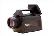
FLIR Systems has launched its new X6580sc thermal imaging camera as a solution for researchers and scientists that require ultra-fast frame rate acquisition of extremely dynamic thermal events.
The versatile FLIR X6580sc camera uniquely ombines high speed and high resolution with ease of use and the flexibility to be configured for just about any scientific or research application. The X6580sc features a high-speed 640x512 digital InSb detector with broadband (1.5-5.5µm) spectral sensitivity and F/3 aperture. The X6580sc provides images up to 350Hz in full frame and up to 4500 Hz in a 320x8 sub-windowing mode....
Specialised Imaging Ltd. has recently delivered a 16-channel SIM-D framing camera capable of capturing images at 1,000,000,000 frames per second, with gating down to 3ns, to the UK’s National Physical Laboratory, which is located in Teddington, Middlesex.
The National Physical Laboratory is the UK’s National Measurement Institute, and is a world-leading centre of excellence in developing and applying the most accurate measurement standards, science and technology available. Gianluca Memoli, Senior Research Scientist at NPL, who is using the SIMD-16 for his cavitation research, says: “I particularly like the possibility of adjusting the timing of each single frame without paying with pixel resolution,....
FLIR Systems has expanded its range of highly affordable, entry level R&D grade thermal imaging kits for academic teaching and industrial research labs.
New additions to the FLIR® portfolio of R&D-grade thermal imaging kits are the portable (< 1kg) and easy-to-operate E40 and T420 entry-level packages. Combining a high performance thermal imaging camera, advanced optics and versatile software the new camera kits bring research grade thermal imaging within the reach of almost any organisation. Unlike thermocouples, spot pyrometers and IR thermometers, the FLIR E40 / T420 thermal imaging kits eliminate risky guesswork with instant non-contact readings that deliver up to 76,800 repeatable, accurate temperature measurements in each thermographic image. Visualizing temperatures from -20°C to +650°C, the FLIR E40 / T420 thermal imaging kits deliver true R&D-grade performance and their high accuracy (±2%) and sensitivity (up to <0.045°C) lets you see the finest thermal variations...
JPK Instruments, a world-leading manufacturer of nanoanalytic instrumentation for research in life sciences and soft matter, reports on how AFM and advanced fluorescence microscopy is being applied in the study of biological membranes in the Centre de Biochimie Structurale (CBS, CNRS and INSERM affiliated) in Montpellier (France).
The CBS includes a research group focused on single molecule physics. Dr Pierre-Emmanuel Milhiet runs a team which applies AFM and advanced fluorescence microscopies (single molecule tracking and single-molecule localization microscopy or SMLM) in the study of both structure and dynamics of biological membranes. Speaking about his work, Dr Milhiet says "One of our aims is to decipher the molecular mechanisms involved in the lateral segregation of membrane components using artificial bilayers and intact cell membranes. Part of our activities is also to develop new methodologies and we have recently mounted a new setup combining a JPK AFM and home-made SMLM (especially PALM and STORM)...
The CVC AN series of colour cameras with crosshairs from STEMMMER IMAGING now offer HD imaging capabilities for medical and scientific imaging applications.
These new versions provide a cost effective way of displaying HD images directly on a monitor without the need for a computer. Cameras are now available in 1920 x 1080 (CVC AN-1080) or 1280 x 720 (CVC AN-072) pixel resolution, both providing 60 frames/s output. The CVC AN-1080 offers the additional benefit of being able to capture images to an SD memory card inserted in the camera back using a wired remote control. The cameras are compatible with C-mount lenses and offer an excellent high definition alternative to standard video cameras...
Anasys Instruments reports on EPFL's publication in Plant Cell on the use of nanoIR to look into the process of photosynthesis to shed more light on how plants produce energy
École Polytechnique Federale de Lausanne, better known as EPFL, has recently reported on how a group of its scientists have used powerful imaging techniques including nanoIR to support a study which sheds light on photosynthesis. All plants use a form of photosynthesis to produce energy, though not all rely exclusively on it. In higher plants, capturing light takes place in specialized compartments called thylakoids. These are found in cell organelles called chloroplasts, which are the equivalent of a power station for the plant....
The telescope door of NASA’s Interface Region Imaging Spectrograph (IRIS) equipped with HORIBA Jobin Yvon diffraction gratings was opened on July 17, 2013.
The IRIS spectrograph has begun to observe with unprecedented detail the lowest parts of the sun's atmosphere, known as the interface region. “The quality of images and spectra we are receiving from IRIS is amazing.” said Dr. Alan Title, IRIS principal investigator. IRIS data will allow scientists to study and better understand the energy transport on the sun. The diffraction gratings for the IRIS spectrograph have been produced by HORIBA Jobin Yvon S.A.S.Longjumeau – France...
Surgeons Benefit from Easy 3D Recording and Video Editing, More Space to Maneuver in the Operating Room and an Upgradable Platform for Future Surgical Guidance Applications
The first 3D surgical microscopes from Leica Microsystems with TrueVision 3D technology inside are available to customers. By incorporating the digital smart 3D system inside select models of Leica Microsystems' surgical microscopes, the two companies have eliminated the need for a separate 3D cart. Surgeons can control the 3D recording functions, without interrupting their workflow, via the microscope hand and foot controls, and OR staff will benefit from easier, faster setup and more space to maneuver in the operating room....
Banook Central Imaging selected by Servier to provide innovative image analysis services based on a combination of RECIST, IrRC and TGR criteria
Banook Central Imaging (Banook CI), a global imaging core lab for drug and device clinical trials, announces today that it has been selected by Servier to assess the potential anti-tumor activity of a novel monoclonal antibody based on the RECIST 1.1 (Response Evaluation Criteria in Solid Tumors) and Immune-Related Response criteria (IrRC)....


 The location of the site where autophagosomal membranes are created within the cell has been the subject of two competing models for many years. Now, a group of researchers from Osaka University led by Prof. Maho Hamasaki has located the origin of autophagosomes within the cell and shown that neither model is correct. Writing in Nature, they show that autophagosomes form at the contact site of the endoplasmic reticulum (ER) with mitochondria. Live cell imaging studies, using an Andor Revolution XD spinning disk confocal microscope equipped with three Andor iXon 3 EMCCD cameras, captured previously hidden detail...

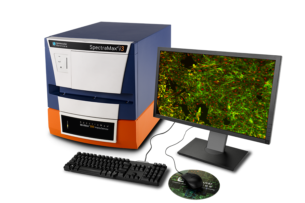 Molecular Devices®, the market leader in microplate reader technology for over 25 years, has announced today the next-generation SpectraMax® MiniMax™ 300 Imaging Cytometer. This MiniMax 300 imaging cytometer enables both cellular visualization and first-of- its-kind stain free cell-based analysis on the field-upgradable SpectraMax® i3 Multi-Mode Microplate Reader. Brightfield and fluorescence based green and red channel cellular image acquisition and analysis is made simple using the SoftMax Pro® software workflow....
 Sorlox Corporation is a pioneer in the development of compact pulsed-plasma devices for use as highly efficient, environmentally friendly power production or for medical isotope production. Using Deuterium as its fuel source - the Sorlox Nautilus Compressor employs an electric field to generate a hot plasma. Using a magnetic field, the Nautilus system compresses the plasma to a high energy state, facilitating fusion and releasing heat that can be used for power generation...
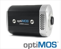 optiMOS is suited for a broad range of fluorescence microscopy applications. The camera is ideal for cell biologists using live cell, multicolor fluorescence; biophysicists studying membrane dynamics and protein and lipid trafficking; as well as neuroscientists looking at ion transport such as electrophysiology, calcium imaging and ratiometric imaging.

 TissueMark software automatically marks and detects potentially cancerous sections in tissue samples with unprecedented levels of speed and accuracy. Technology has the potential to save pathologists a combined 250,000 working days of effort per annum. Leap in precision equates to the shift from a freehand outline sketch of a country’s border to a high resolution map of the whole country. New technology, developed in Belfast, will soon enable pathologists to automate the process of marking tissue samples with unprecedented accuracy. TissueMark, developed by digital pathology specialists PathXL, analyses the detailed structural patterns in tissue samples and marks the boundaries of potentially cancerous sections for more detailed analysis...
 The product is available in a 1 L bottle for Coplin jars or staining dishes and a 4 L container with flow control spout for filling baths on the Little Dipper Processor and VP 2000 instruments. No dilution is required and the reagent can be stored at room temperature. The product joins an expanding line of FISH slide processing products from SciGene including FISH Wash Buffers, CytoZyme Stabilized Pepsin and CytoBond Removable Coverslip Sealant...

 The versatile FLIR X6580sc camera uniquely ombines high speed and high resolution with ease of use and the flexibility to be configured for just about any scientific or research application. The X6580sc features a high-speed 640x512 digital InSb detector with broadband (1.5-5.5µm) spectral sensitivity and F/3 aperture. The X6580sc provides images up to 350Hz in full frame and up to 4500 Hz in a 320x8 sub-windowing mode....

 The CBS includes a research group focused on single molecule physics. Dr Pierre-Emmanuel Milhiet runs a team which applies AFM and advanced fluorescence microscopies (single molecule tracking and single-molecule localization microscopy or SMLM) in the study of both structure and dynamics of biological membranes. Speaking about his work, Dr Milhiet says "One of our aims is to decipher the molecular mechanisms involved in the lateral segregation of membrane components using artificial bilayers and intact cell membranes. Part of our activities is also to develop new methodologies and we have recently mounted a new setup combining a JPK AFM and home-made SMLM (especially PALM and STORM)...
 These new versions provide a cost effective way of displaying HD images directly on a monitor without the need for a computer. Cameras are now available in 1920 x 1080 (CVC AN-1080) or 1280 x 720 (CVC AN-072) pixel resolution, both providing 60 frames/s output. The CVC AN-1080 offers the additional benefit of being able to capture images to an SD memory card inserted in the camera back using a wired remote control. The cameras are compatible with C-mount lenses and offer an excellent high definition alternative to standard video cameras...
 École Polytechnique Federale de Lausanne, better known as EPFL, has recently reported on how a group of its scientists have used powerful imaging techniques including nanoIR to support a study which sheds light on photosynthesis. All plants use a form of photosynthesis to produce energy, though not all rely exclusively on it. In higher plants, capturing light takes place in specialized compartments called thylakoids. These are found in cell organelles called chloroplasts, which are the equivalent of a power station for the plant....
 The IRIS spectrograph has begun to observe with unprecedented detail the lowest parts of the sun's atmosphere, known as the interface region. “The quality of images and spectra we are receiving from IRIS is amazing.” said Dr. Alan Title, IRIS principal investigator. IRIS data will allow scientists to study and better understand the energy transport on the sun. The diffraction gratings for the IRIS spectrograph have been produced by HORIBA Jobin Yvon S.A.S.Longjumeau – France...
 The first 3D surgical microscopes from Leica Microsystems with TrueVision 3D technology inside are available to customers. By incorporating the digital smart 3D system inside select models of Leica Microsystems' surgical microscopes, the two companies have eliminated the need for a separate 3D cart. Surgeons can control the 3D recording functions, without interrupting their workflow, via the microscope hand and foot controls, and OR staff will benefit from easier, faster setup and more space to maneuver in the operating room....