We bring you more exciting news about Photonex London Roadshow.
The programme has now been finalised with an excellent calibre of speakers set to give presentations. Attend this event to broaden your knowledge in the world of photonics by attending a key meeting for photonics technologies in the life sciences. The technical committee have commented that this international quality 'invited' programme includes presentations by eminent and leading European research scientists...
FLIR Systems has introduced the new FLIR X6900sc as the world's fastest 640 x 512 pixel resolution thermal camera for high-speed science applications.
The new thermal imager is designed to record 1000 fps at full resolution onto the camera's RAM for up to 26 seconds. Whether measuring temperatures on fast moving objects or characterizing the thermal transient of objects as they heat up, this new camera offers the rapid frame speed, high resolution, and thermal sensitivity required to virtually stop motion enabling accurate temperature readings, and recording of gradients across...
MR Solutions’ new 3D in vivo confocal microscope for use in preclinical research provides a magnification range of up to 1000 times, allowing researchers to examine cellular details within a live small animal eliminating the need for a surgical biopsy - saving time and substantially reducing costs.
MR Solutions, the world leader in preclinical scanning, has partnered with Optiscan1 to introduce CellLiveTM, their second generation endomicroscopy platform to the preclinical market. The handheld Optiscan probe which is less than 3.6 mm in diameter delivers micron – smaller than a millionth of a metre - resolution. The Optiscan uses a fluorescence2, confocal3 imaging system to achieve this...
The University of Tokyo's Department of Mechanical Engineering was established in 1879, providing education based on four disciplines; mechanics, materials, hydrodynamics and thermodynamics.
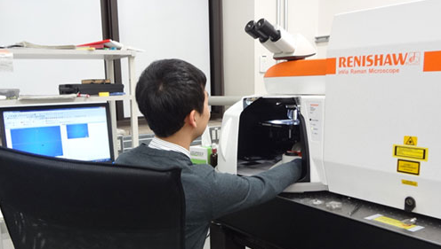
Within the Department, the Maruyama-Chiashi Laboratory focuses its research on the synthesis and analysis of carbon nanotubes (CNT), graphene and other nano-materials. They study applications related to the development of energy related devices, such as solar cells. The laboratory uses scanning Raman spectroscopy as an important tool for the investigation of the synthesized materials and their structure...
JPK Instruments, a world-leading manufacturer of nanoanalytic instrumentation for research in life sciences and soft matter, reports on the use of their NanoTracker™ 2 optical tweezers system which is being used to study the physical and chemical properties of micro-bubbles in the Department of Mechanical Engineering at the Shibaura Institute of Technology under the leadership of Associate Professor, Dr Yoko Yamanishi.
Dr Yoko Yamanishi is an Associate Professor in the Department of Mechanical Engineering at Shibaura Institute of Technology, Tokyo, Japan. She leads the Yamanishi Laboratory - the Micro-nano Functional Fluid Laboratory. The Laboratory's goals aim to clarify unknown function of cells by using micro-nano technology based on mechanical, electrical and bio-medical engineering. It targets a contribution to the development of cellular scale...
Prior Scientific has announced the FB201 co-axial coarse and fine focusing block, designed for OEM microscopy applications where precise fine focus adjustment is required.
Rack and pinion mounting allows the FB201 to produce smooth, precise focusing over 29 mm of travel. A large coarse focus mechanism incorporates a slip clutch and tension adjustment. Fine focus adjustment control is graduated in two micron divisions throughout the coarse focus range. The focus block can be easily adapted to various mounting configurations and can support up to 5 kg. Motorised variants able to...
At last, an affordable solution for the scanning of histology and Cytology microscope slides.
The new Slide Scanner available from Eikonix now brings high quality slide scanning to a much wider market. This compact device utilises a host of the latest automated imaging technologies with a proven scanning engine to create an effective and reliable tool for the rapid acquisition of whole slide images from standard glass microscope slides. The easy to use system can be operated by anyone and requires no specialist training. The intuitive software effortlessly guides the user through each step to produce images of exceptional quality.
Fluorescence analysis workstations are one of many products manufactured by Spectronics Corporation to aid laboratory technicians.
Built to exacting standards, they’re used for viewing and analyzing fluorescent samples with both epi-illumination and trans-illumination light sources. The Spectroline® CM-10MP mini viewing cabinet is perfect for any type of laboratory application requiring high-contrast fluorescence analysis. It’s also excellent for viewing TLC plates and for quality control inspection of PC boards. The CM-10MP is an “all-in-one tool” for the laboratory technician...
Combines Accuracy with Affordability to Deliver Rapid Count Results
Synbiosis, a long-established, expert manufacturer of automated microbiological systems, is delighted to introduce the aCOLyte 3 HD its next generation, automated colony counter for microbiologists that demand a sensitive, budget system to quickly and accurately count colonies of all sizes and colours. The new aCOLyte 3 HD colony counter, features a high resolution megapixel CCD camera for accurate detection of different coloured...
Union Biometrica develops tools for model organism research.
The NEW VAST BioImager platform™ (Vertebrate Automated Screening Technology) is designed for zebrafish researchers who need to image large numbers of 2–7 day old sedated zebrafish larvae. VAST automation avoids the time consuming and tedious steps of manual manipulation of each larva during screens. VAST loads each larva into a capillary and then rotates it 360 degrees to determine the orientation of the fish...
ZEISS introduced two new digital microscope cameras during the annual meeting of the American Society for Cell Biology (ASCB 2015) in San Diego.
ZEISS Axiocam 702 mono and ZEISS Axiocam 512 color complement the current portfolio of high-speed USB 3.0 microscope cameras. With ZEISS Axiocam 702 mono ZEISS for the first time introduces a microscope camera with a scientific CMOS sensor. Users benefit from low read noise, excellent low light sensitivity and high speed for live cell imaging and acquisition of fast processes. ZEISS Axiocam 702 mono features a 1/1.2" (13.3 mm diagonal)...
LED fluorescence illumination devices that provide a simple solution for microscope users wishing to upgrade their microscope for fluorescence applications.
New from Eikonix, the imaging specialists, is a range of LED fluorescence illumination devices that provide a simple solution for microscope users wishing to upgrade their microscope for fluorescence applications. The MF-LED module which has built-in coloured LED’s can easily upgrade a traditional infinity upright microscope to give fluorescence functionality. It is optically compatible with many popular microscope brands such as Olympus, Nikon, Leica, Zeiss and others....
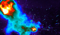
FLIR Systems announces a new addition to its RS-Series of long-range infrared camera systems designed for range tracking, target signature, research, and scientific applications.
The new FLIR RS8300 couples a proprietary high-speed HD MWIR detector with a 10x continuous zoom lens in a sealed, ruggedized enclosure. Combining reliability with leading edge performance, the FLIR RS8300 delivers stunning megapixel infrared imagery at up to 200 megapixels per second. To capture the most fleeting of events the camera is capable of lightning fast frame rates from full-frame resolution 14-bit...
Broadening the capability of the most versatile 3D X-ray microscopy instrument
The new ZEISS FPX flat panel extension for the ZEISS Xradia Versa 500-series of 3D X-ray microscopes delivers large-sample, high throughput scanning with best-in-class image quality. Combined with the high resolution of ZEISS Xradia Versa X-ray microscopes (XRM), the new ZEISS FPX enhances imaging flexibility and creates workflow efficiencies with an all-in-one system for industrial development and academic research...
Leica Biosystems, a global leader in anatomic pathology solutions and workflow automation, and Advanced Cell Diagnostics, Inc. (ACD), a world leader in RNA biomarker analysis for precision medicine, today announced a comprehensive partnership to develop and commercialize tissue-based diagnostic tests based on ACD's RNAscope in situ hybridization (ISH) assays on Leica Biosystems' BOND clinical advanced staining instruments.
The agreement supports Leica Biosystems' development and commercialization of fully automated RNAscope-based companion diagnostic (CDx) tests in partnership with biopharmaceutical companies. The combination of ACD's RNAscope technology with Leica Biosystems' fully automated pathology solutions allows companies and laboratories in clinical markets to integrate the power of new RNA-based biomarker tests into the...
The new ZEISS Smartproof 5 widefield confocal microscope is designed for a wide range of industrial applications in quality assurance and quality control departments, production environments and R&D labs. The microscope system provides 3D reconstructions and roughness measurements for a wide range of work piece surfaces. Users benefit from an integrated design, repeatable results and high throughput...
The Omega Lum G from Aplegen is rapidly growing into the gel and western blot imaging documentation system of choice for many people. Installations are growing across Europe as more and more researchers learn about the high performance and yet low cost for this very popular system.
As with all Aplegen systems it is powered by the intelligent SmartCapture Technology which simplifies imaging to the point where the user just has to pick an application and the image is automatically captured with the optimum exposure. This automatically controlled system even takes care of filter selection and focusing. Within seconds the user will have a high quality publication quality image at the click of a button....
Prior Scientific reports that its OptiScan® controller together with associated microscope stage and focus motor have been used to create stunning artistic designs.
Using this equipment, Richard Weston, a former Professor of Architecture at Cardiff University (UK) has acquired high magnification images of minerals, such as agate, fluorite, and calcite, to produce unique designs for a range of products including scarves, glass panels, and in one case a facade for a three story house. Asked about his novel use of the Prior stage and stepper motor, Richard Weston commented...
Denatures and Hybridizes 72 Slides in Half the Cost and Bench Space of SIX ThermoBrite Instruments
A single CytoBrite Slide Incubation System can perform the short denaturation step on multiple batches of up to 12 FISH slides with each batch then transferred to the CytoBrite Slide Oven for overnight incubation. The combined instruments hybridize up to 72 FISH slides simultaneously; the equivalent capacity of SIX ThermoBrite instruments at less than half the cost and bench space. Removable slide trays streamline work by holding...
Deben, a leading provider of in-situ testing stages together with innovative accessories and components for electron microscopy, reports on the use of a Microtest tensile stage in the Institute of Photonics & Quantum Sciences at Heriot-Watt University, Edinburgh.
It is being used for stress analysis studies of ceramics and engineering plastics. Dr Peter Schemmel is a postdoctoral researcher in Applied Photonics at Heriot-Watt University. He is part of the team at the Institute of Photonics and Quantum Sciences in a group which focuses on stress analysis in ceramics and engineering plastics. They are particularly interested in ceramic thermal barrier coatings used on aeroplane engine turbine blades or...
The LumaSpec LS800S from Prior Scientific is a compact visible light spectrophotometer which provides quantitative spectral power data enabling users to characterise and monitor the illumination source of their microscope system.
Operating from 350 nm to 800 nm with 1.5 nm resolution, the LumaSpec LS800S uses an illumination target slide mounted on your microscope to provide accurate and precise information about the illumination in the sample plane, leading to the most relevant information being obtained. The LumaSpec LS800S is capable of analysing the output from a wide variety of light sources, ranging from the dimmest LED to a powerful mercury...
Presenting a faster and more efficient alternative to electron microscopy, digital light microscopy has been advancing investigations into the mechanical properties of bone, as explored in a new application note from Olympus.
Unravelling the mechanical properties of bone with micro-indentation testing has significance in both health and disease. At the University Medical Center Hamburg-Eppendorf, Dr. Björn Busse and his research group have advanced their micro-indentation testing technique using an Olympus industrial inspection digital light microscope, as an alternative to electron microscopy. The cutting edge research is described in a new application...


 The new thermal imager is designed to record 1000 fps at full resolution onto the camera's RAM for up to 26 seconds. Whether measuring temperatures on fast moving objects or characterizing the thermal transient of objects as they heat up, this new camera offers the rapid frame speed, high resolution, and thermal sensitivity required to virtually stop motion enabling accurate temperature readings, and recording of gradients across...
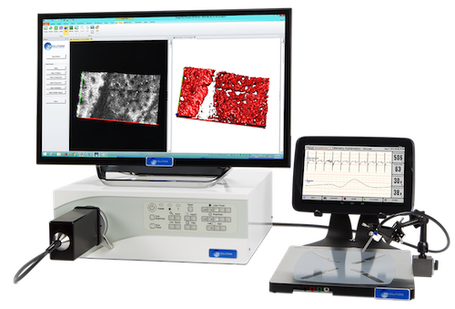
 Within the Department, the Maruyama-Chiashi Laboratory focuses its research on the synthesis and analysis of carbon nanotubes (CNT), graphene and other nano-materials. They study applications related to the development of energy related devices, such as solar cells. The laboratory uses scanning Raman spectroscopy as an important tool for the investigation of the synthesized materials and their structure...
 Dr Yoko Yamanishi is an Associate Professor in the Department of Mechanical Engineering at Shibaura Institute of Technology, Tokyo, Japan. She leads the Yamanishi Laboratory - the Micro-nano Functional Fluid Laboratory. The Laboratory's goals aim to clarify unknown function of cells by using micro-nano technology based on mechanical, electrical and bio-medical engineering. It targets a contribution to the development of cellular scale...
 Rack and pinion mounting allows the FB201 to produce smooth, precise focusing over 29 mm of travel. A large coarse focus mechanism incorporates a slip clutch and tension adjustment. Fine focus adjustment control is graduated in two micron divisions throughout the coarse focus range. The focus block can be easily adapted to various mounting configurations and can support up to 5 kg. Motorised variants able to...
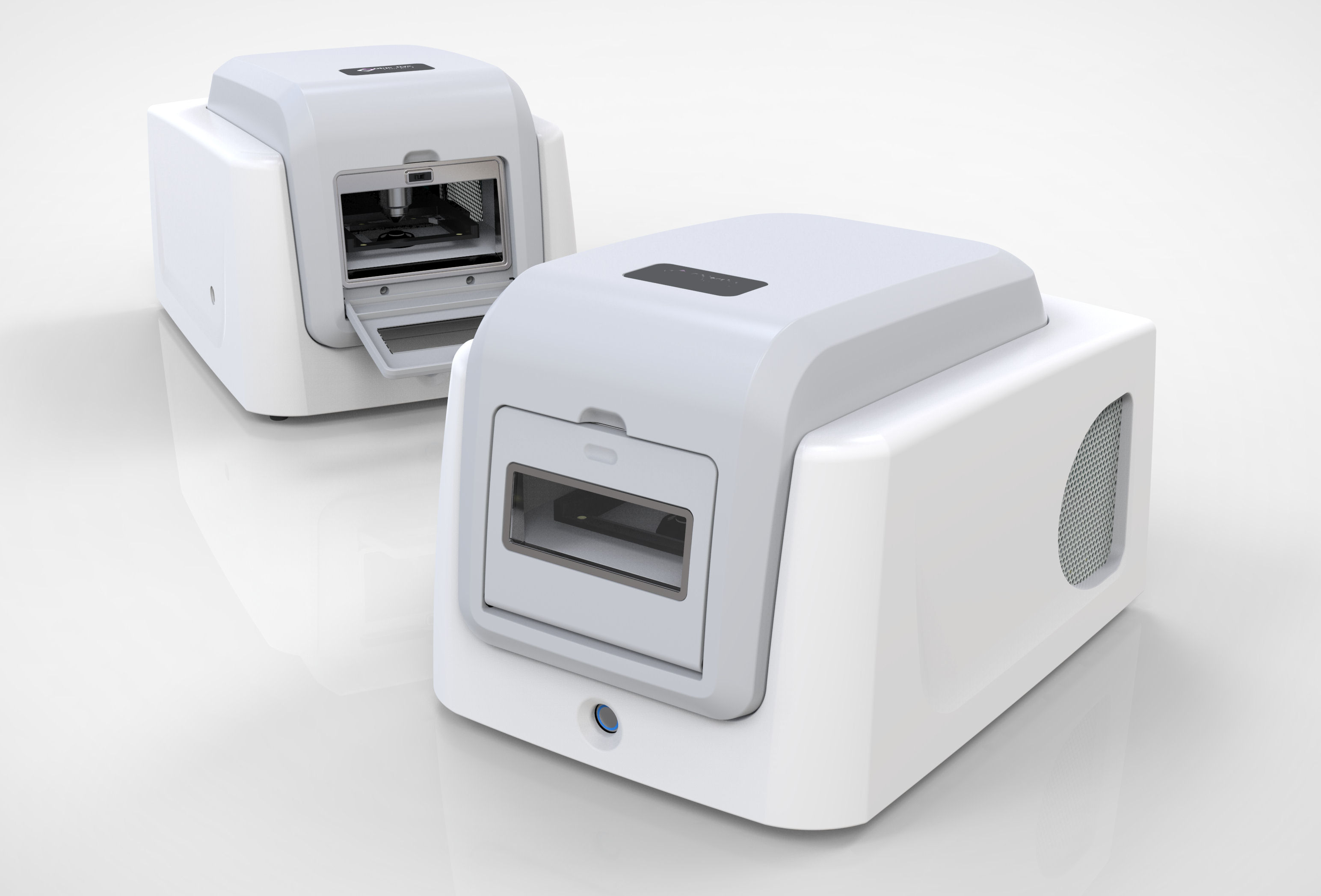 The new Slide Scanner available from Eikonix now brings high quality slide scanning to a much wider market. This compact device utilises a host of the latest automated imaging technologies with a proven scanning engine to create an effective and reliable tool for the rapid acquisition of whole slide images from standard glass microscope slides. The easy to use system can be operated by anyone and requires no specialist training. The intuitive software effortlessly guides the user through each step to produce images of exceptional quality.
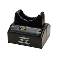 Built to exacting standards, they’re used for viewing and analyzing fluorescent samples with both epi-illumination and trans-illumination light sources. The Spectroline® CM-10MP mini viewing cabinet is perfect for any type of laboratory application requiring high-contrast fluorescence analysis. It’s also excellent for viewing TLC plates and for quality control inspection of PC boards. The CM-10MP is an “all-in-one tool” for the laboratory technician...
 Synbiosis, a long-established, expert manufacturer of automated microbiological systems, is delighted to introduce the aCOLyte 3 HD its next generation, automated colony counter for microbiologists that demand a sensitive, budget system to quickly and accurately count colonies of all sizes and colours. The new aCOLyte 3 HD colony counter, features a high resolution megapixel CCD camera for accurate detection of different coloured...
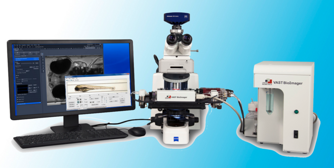 The NEW VAST BioImager platform™ (Vertebrate Automated Screening Technology) is designed for zebrafish researchers who need to image large numbers of 2–7 day old sedated zebrafish larvae. VAST automation avoids the time consuming and tedious steps of manual manipulation of each larva during screens. VAST loads each larva into a capillary and then rotates it 360 degrees to determine the orientation of the fish...
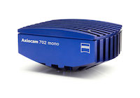 ZEISS Axiocam 702 mono and ZEISS Axiocam 512 color complement the current portfolio of high-speed USB 3.0 microscope cameras. With ZEISS Axiocam 702 mono ZEISS for the first time introduces a microscope camera with a scientific CMOS sensor. Users benefit from low read noise, excellent low light sensitivity and high speed for live cell imaging and acquisition of fast processes. ZEISS Axiocam 702 mono features a 1/1.2" (13.3 mm diagonal)...
 New from Eikonix, the imaging specialists, is a range of LED fluorescence illumination devices that provide a simple solution for microscope users wishing to upgrade their microscope for fluorescence applications. The MF-LED module which has built-in coloured LED’s can easily upgrade a traditional infinity upright microscope to give fluorescence functionality. It is optically compatible with many popular microscope brands such as Olympus, Nikon, Leica, Zeiss and others....
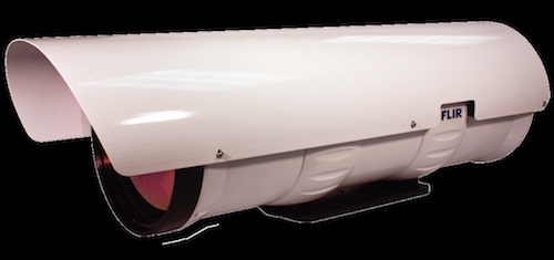 The new FLIR RS8300 couples a proprietary high-speed HD MWIR detector with a 10x continuous zoom lens in a sealed, ruggedized enclosure. Combining reliability with leading edge performance, the FLIR RS8300 delivers stunning megapixel infrared imagery at up to 200 megapixels per second. To capture the most fleeting of events the camera is capable of lightning fast frame rates from full-frame resolution 14-bit...
 The new ZEISS FPX flat panel extension for the ZEISS Xradia Versa 500-series of 3D X-ray microscopes delivers large-sample, high throughput scanning with best-in-class image quality. Combined with the high resolution of ZEISS Xradia Versa X-ray microscopes (XRM), the new ZEISS FPX enhances imaging flexibility and creates workflow efficiencies with an all-in-one system for industrial development and academic research...
 The agreement supports Leica Biosystems' development and commercialization of fully automated RNAscope-based companion diagnostic (CDx) tests in partnership with biopharmaceutical companies. The combination of ACD's RNAscope technology with Leica Biosystems' fully automated pathology solutions allows companies and laboratories in clinical markets to integrate the power of new RNA-based biomarker tests into the...
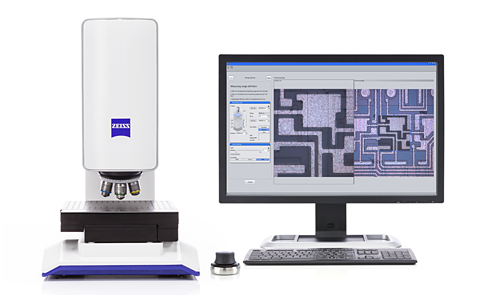 The new ZEISS Smartproof 5 widefield confocal microscope is designed for a wide range of industrial applications in quality assurance and quality control departments, production environments and R&D labs. The microscope system provides 3D reconstructions and roughness measurements for a wide range of work piece surfaces. Users benefit from an integrated design, repeatable results and high throughput...
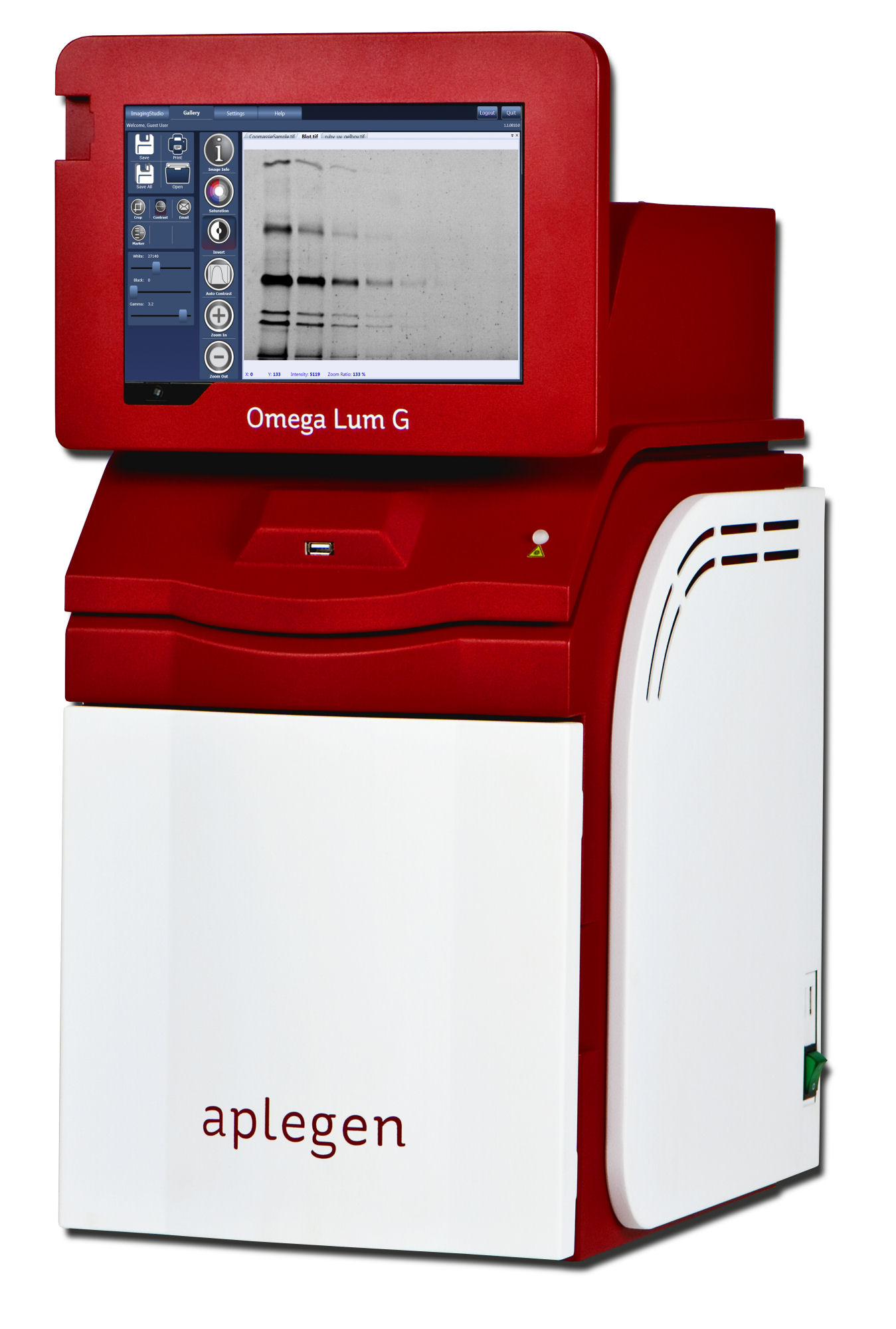 As with all Aplegen systems it is powered by the intelligent SmartCapture Technology which simplifies imaging to the point where the user just has to pick an application and the image is automatically captured with the optimum exposure. This automatically controlled system even takes care of filter selection and focusing. Within seconds the user will have a high quality publication quality image at the click of a button....
 Using this equipment, Richard Weston, a former Professor of Architecture at Cardiff University (UK) has acquired high magnification images of minerals, such as agate, fluorite, and calcite, to produce unique designs for a range of products including scarves, glass panels, and in one case a facade for a three story house.
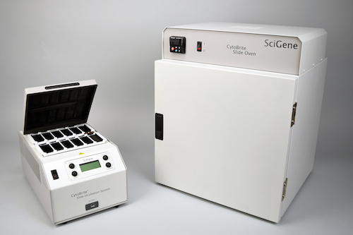
 It is being used for stress analysis studies of ceramics and engineering plastics.
  Operating from 350 nm to 800 nm with 1.5 nm resolution, the LumaSpec LS800S uses an illumination target slide mounted on your microscope to provide accurate and precise information about the illumination in the sample plane, leading to the most relevant information being obtained.
 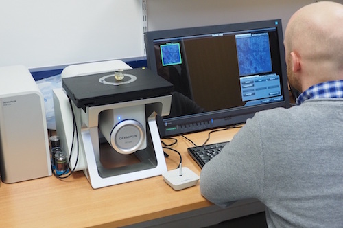 Unravelling the mechanical properties of bone with micro-indentation testing has significance in both health and disease. At the University Medical Center Hamburg-Eppendorf, Dr. Björn Busse and his research group have advanced their micro-indentation testing technique using an