
Channels
Special Offers & Promotions
New diagnostic technique improves breast cancer screening
Flexible coils for magnetic resonance imaging are being designed to search for breast cancer faster and with less disadvantages. In a project funded by the Austrian Science Fund FWF, a prototype is now being developed in Vienna.
Breast cancer is among the most common forms of cancer and is the most frequent type found among women in the western world, making early detection particularly important. Imaging examinations of the female breast, referred to as “mammography”, include X-ray methods or, alternatively, less risky but also more expensive, magnetic resonance imaging (MRI). MRI exposes the human body to a static and a high-frequency magnetic field, which electromagnetically excites the nuclei of the hydrogen atoms in the body’s tissue. This reaction is then used to record a three-dimensional image from inside the body. An MRI examination does, however, present some practical disadvantages. In an international project funded by the FWF together with the French funding agency ANR, physicist Elmar Laistler is now working on making this procedure for detecting breast cancer less cumbersome and cheaper.
No X-ray risk
“One advantage of examining the breast with MRI is the avoidance of X-rays, as used in classical mammography,” says Laistler. The risks involved in such ionising radiation are of concern, in particular for young women. “Further advantages of MRI are significantly higher sensitivity and better resolution of the measurement.” According to Laistler, MRI examination is well on its way to becoming the standard method, but some issues remain: “Above all, it is expensive and takes longer.” This is why he and his team want to bring about improvements to the hardware.
“So far, MR mammography has involved having women lie face down in the MR scanner tube,” explains the medical physicist. “In MRI, the actual image is always recorded with radio frequency coils.” These coils are housed in two one-size-fits-all “cups”. The patient lies on her stomach so that her breasts settle into these cups. “This does not work equally well for all women and all breast sizes, because the coils are more efficient and give better results depending on whether they are a good fit for the respective body shape,” explains Laistler.
Examination lying face up
The researcher wants to turn this situation around: “In our case, the woman lies face up and the coils should be flexible enough to be put on like a waistcoat.” This approach has several advantages. “An essential aspect of the project is that lying on the back results in the breast being flattened, which means a much larger part of it is close to the receiver,” notes Laistler. “In this way, the signal is stronger and the measuring time can be shortened” – regardless of the size of the breast.
In addition, a biopsy may often be required after the MRI examination, where tissue suspected of being a tumour is removed with a needle. “That’s the gold standard,” Laistler reports. During the biopsy, the patient usually lies on her back or on her side. “It is often difficult for the doctor to find the spot he or she has seen on the downward-facing MR image because the breast is of a different shape depending on the posture. With our method, the shape doesn’t change much.”
Correcting for breathing motion
There is, however, one catch here: the patient’s breathing. Raising and lowering of the chest distorts the image and needs to be factored out computationally. “That’s why we’ve added motion sensors to our waistcoat. Together with a correction algorithm, this remedies the situation and delivers sharp images,” says the researcher, describing the process.
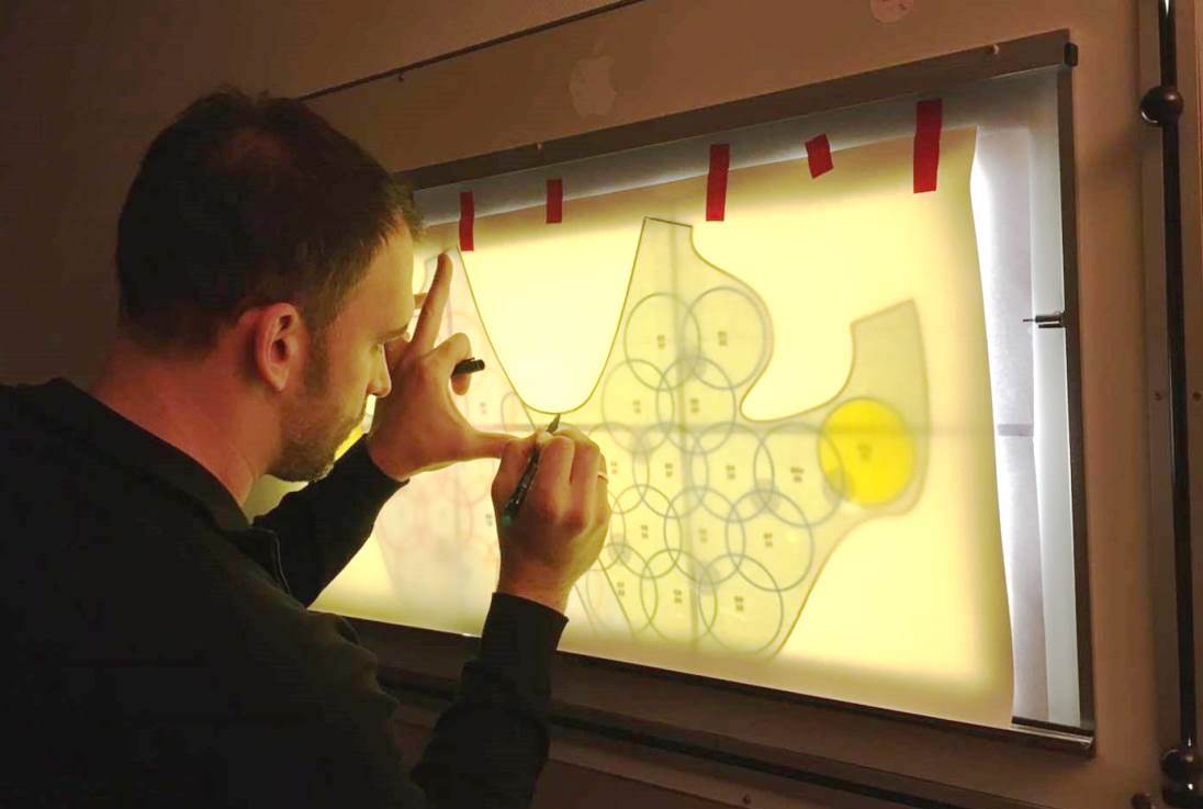
A prototype of this waistcoat is currently being made. It will contain 32 coils, each with a diameter of eight centimetres, consisting of coaxial cables. A receiver unit at the end of the cable feeds the signal into the computer. Laistler’s group in Vienna is concentrating on the hardware development part of the project. Motion sensors and correction software are being developed by a group at the Université de Lorraine in Nancy. The know-how relating to the coils is the result of cooperation with Paris-Sud University, where Laistler completed the first year of his doctorate. An Associate Professor at the Medical University of Vienna since 2018, Elmar Laistler emphasises how important projects like this are for a young scientist in building up a research group. His group has had approximately eight members over several years, most of whom are financed through FWF projects.
A medical device at the end of the road
“At the end of the project, we expect there to be a complete hard- and software package for MRI breast examinations,” says Laistler. If this goal is achieved, the prototype will then be developed into a medical device in cooperation with partners from industry – an outcome the researcher has already successfully demonstrated in previous projects.
Personal details
Elmar Laistler is an Associate Professor at the Center for Medical Physics and Biomedical Engineering at the Medical University of Vienna. The physicist is the head of the group for radio frequency physics, and his research interest includes hardware and software for high-field magnetic resonance investigations.
Publications
Frass-Kriegl R, Navarro de Lara LI, Pichler M, Sieg J, Moser E, Windischberger C, Laistler E.: Flexible 23-channel coil array for high-resolution magnetic resonance imaging at 3 Tesla, in: PLoS ONE 2018
Hosseinnezhadian S, Frass-Kriegl R, Goluch-Roat S, Pichler M, Sieg J, Vít M, Poirier-Quinot M, Darrasse L, Moser E, Ginefri J-C, Laistler E.: A flexible 12-channel transceiver array of transmission line resonators for 7 T MRI, in: Journal of Magnetic Resonance 2018
Goluch S, Frass-Kriegl R, Meyerspeer M, Pichler M, Sieg J, Gajdošík M, Krššák M, Laistler E.: Proton-decoupled carbon magnetic resonance spectroscopy in human calf muscles at 7 T using a multi-channel radiofrequency coil, in: Scientific Reports 2018
News Channels
- Latest News
- New Laboratory Products
- Industry News
- Laboratory Automation | IT Solutions
- Microscopy | Image Analysis
- Separation Science
- Research | Case Studies
- Video Presentations
- Events | Conferences
Subscribe to any of our newsletters for the latest on new laboratory products, industry news, case studies and much more!

Popular this Month
Top 10 most popular articles this month
Today's Picks
Looking for a Supplier?
Search by company or by product
Please note Lab Bulletin does not sell, supply any of the products featured on this website. If you have an enquiry, please use the contact form below the article or company profile and we will send your request to the supplier so that they can contact you directly.
Lab Bulletin is published by newleaf marketing communications ltd.
Media Partners