

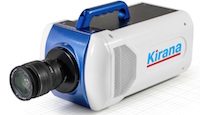
 The Specialised Imaging Kirana camera provides REL, Inc with the ability to create high-resolution images of the ultra high-speed events that often occur with materials testing. Taking up to 5,000,000 frames per second, and shuttering every 100 nanoseconds, there is no compromise in capturing high quality data. The data is exported in 180 frames so each test can be easily saved and viewed. The initial applications undertaken...
 Uniquely combining high performance, leading edge features and affordability the omniDOC series provide an easy-to-use, yet powerful gel imaging system that satisfies the needs of most laboratories. The omniDOCi shares all the same features as the standard omniDOC but with the added benefit of wireless connectivity enabling the system to be run in a darkroom from a remote PC or tablet....
 Users of the SPARC technology at Chalmers University of Technology in Gothenburg, Sweden are applying cathodoluminescence measurements to study the properties of nanophotonic devices. Dr Ruggero Verre is a post-doctoral researcher in the Department of Applied Physics - Division of Bionanophotonics at the Chalmers University of Technology in Gothenburg in southern Sweden. His work in the group headed by...
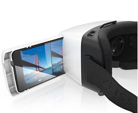 The attention of the young development team was sharply focused on user-friendliness, design and technology. To dive into virtual worlds, all the user needs to do is select an appropriate VR app and slide his or her smartphone into the headset using the tray provided. ZEISS VR ONE will initially be available worldwide with a tray for the Samsung Galaxy S5 smartphone and the iPhone 6. Other models are in the pipeline. The Computer Aided...
 ZEISS and the Howard Hughes Medical Institute’s Janelia Research Campus had signed an exclusive license agreement for the commercialization of Lattice light sheet microscopy earlier this year. By granting the exclusive sublicense to 3i, ZEISS is contributing to making Lattice light sheet microscopy available to scientists and early adopters in a timely manner. 3i will sell and support the development system based on Dr. Eric Betzig’s original design...
 Heading up this new organization is Dr Stefan Kaemmer who has been appointed General Manager of US Operations. Respected German technology leaders, JPK Instruments, are pleased to announce the opening of the first USA-based offices. With more than twelve years supplying nanoscale solutions worldwide for researchers in the bio, life and materials sciences, JPK is to open their offices in Southern California to support users across the...
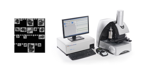 The ability of uncontrolled agglomeration to substantially impact the performance and value of powder products makes efficient agglomerate detection vital across a number of industries. ‘Identification of agglomerates using automated image analysis’ presents practical strategies for efficiently and robustly differentiating agglomerates from primary particles, to support product development, QC and process troubleshooting....
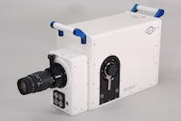 Incorporating a supplementary optical port, that uses a beamsplitter to deliver 50% of the primary image to a secondary image plane, allowing secondary instruments to share the same optical axis, thereby providing true undistorted datasets for simultaneous data measurement. Recording simultaneous ultra fast two-dimensional and time-resolved images is of significant interest to scientists in a growing number of fields of study including...
 The new kit uses imaging flow cytometry to obtain statistically significant quantitative assessment of NF?B translocation, as well as visual identification of the translocation at a single-cell level.
 The Australian Research Council Centre of Excellence in Plant Cell Walls is a collaborative project involving the Universities of Adelaide, Melbourne and Queensland in partnership with South Australia State Government and seven international institutions. Their aim is to advance fundamental scientific understanding of plant cell wall biology. Cell wall composition determines the quality of most plant-based products used in modern human societies...
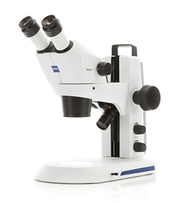 ZEISS is introducing two new compact Greenough stereo microscopes for education, lab routine and industrial inspection: ZEISS Stemi 305 and ZEISS Stemi 508. Users are able to observe their samples in true color, 3D, with high contrast and free of distortion or color fringes. ZEISS Stemi 305 offers a 5:1 zoom. The microscope is easy to use and everything is integrated: LED illumination, reflected and transmitted light illuminations and documentation. Users choose...
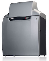 Researchers at the major university are using a G:BOX Chemi XRQ multi-task imager to precisely analyse agarose gels of mouse and rabbit DNA stained with the safe fluorescent dye SybrSafe®. They are also utilising the G:BOX Chemi XRQ to easily image both large and small SDS-PAGE gels and chemiluminescent Western blots of proteins. This is allowing scientists at the university to accurately detect proteins and is contributing to identifying potential therapeutic targets in...
 Peter Friedl holds the chair for Microscopical Imaging of the Cell at the St. Radboud University Nijmegen Medical Centre, University of Nijmegen, The Netherlands. He also holds a 20% joint-appointment as head of the imaging section at the David H. Koch Center, Department of Genitourinary Oncology, MD Anderson Cancer Center, Houston, TX, USA. His research work focuses on imaging cancer growth, metastasis and therapy response in the...
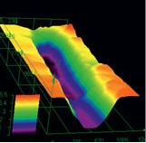 Employing the Olympus DSX100 Opto-digital system, Dr Farrugia’s research at the University of Abertay (Dundee, Scotland) has been greatly enhanced through a combination of digital and optical technologies. Such advanced light microscopy has yielded greater insights into a variety of forensic samples, revealing fingerprints on metal, pen marks on paper and characterising unique designs on seized illicit tablets...
 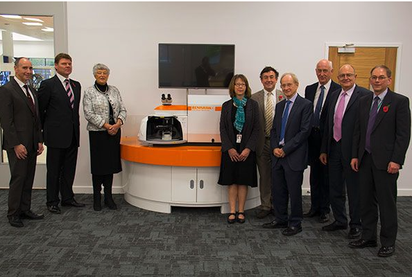 This prestigious award was granted for the continuous development of the company's inVia Raman microscope, with ultra-fast Raman imaging, which enables the rapid generation of high definition 2D and 3D chemical images for material analysis. This Queen's Award for Enterprise 2014, granted in the Innovations category, is particularly special, as it is the first to recognise Renishaw's achievements outside of the field of industrial metrology...
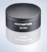 Designed for the convenience of everyday use and ideal under low light conditions, the new 5 megapixel SC50 microscope camera from Olympus is built on the latest CMOS chip technology to deliver improved speed, sensitivity and resolution at a truly affordable price. Achieving Full HD images at 32 fps, high speed imaging with the SC50 captures and records dynamic events. The camera delivers clear, high quality images to nearly every kind of...
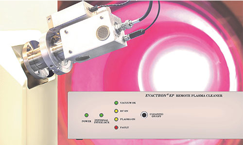 This provides a new approach to plasma cleaning designed for those in the physics and materials science communities building and using custom-designed vacuum systems. The Evactron EP Remote Plasma Source from XEI Scientific uses flowing afterglow cleaning with air to clean carbon compounds from vacuum chambers operating with turbomolecular pumps. The unique new system has instant ignition from any vacuum level...
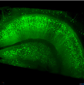 Clearing methods make tissue transparent, thus allowing scientists to image deep into large biological samples such as tissue sections, brains, embryos, organs, spheroids or biopsies. The enhanced optical penetration depth even allows detection of fluorescent signals from whole organs. This means clearing is a promising technique when, for example, investigating neuronal networks in mouse brains....