The all new Nikon SMZ25 and SMZ18 stereomicroscopes have evolved to meet the increasing needs for imaging systems that range from single cells to whole organisms. Featuring a large zoom ratio of 25:1 for the SMZ25, superior resolution and exceptional fluorescence transmission capability, the ergonomic, easy-to-use, fully motorised SMZ25 and manual SMZ18 are ideal for all bioscience applications....
Bruker Corporation today announced that it has signed an exclusive patent license agreement with 3M Company, which allows Bruker to include 3M patented innovations relating to matrix-assisted laser desorption ionization (MALDI) mass spectrometry imaging.
The licensed 3M patents are directed to a technique for performing mass spectrometry analysis on proteins in tissue that has been preserved in paraffin. The technology enables researchers to more easily study formalin-fixed, paraffin-embedded (FFPE) tissue for life-science research and drug development. MALDI imaging has been increasingly used to analyze clinically relevant tissues such as tumor biopsies. The molecular phenotypes observed by MALDI imaging have been shown to correlate with parameters such as disease status or patient outcome, and have been successfully applied to the classification of tissue samples.
JPK Instruments, a world-leading manufacturer of nanoanalytic instrumentation for research in life sciences and soft matter, reports on the work of the Nano-Mechanics Laboratory at the Indian Institute of Science Education & Research (IISER) Pune, India.
The Nano-mechanics group develops and uses novel techniques to understand molecular processes and has recently developed new instrumentation in the fields of single molecule force spectroscopy and correlation spectroscopy. There are three main areas of research: protein folding using AFM, measuring the mechanical properties of supported lipid bilayers and following the mechanical response of nano-confined fluids. This last area is performed on home-built instruments....
Read MoreScientists at CENIMAT and the Materials Science Department, New University of Lisbon, are investigating LCP polymer blends using the Linkam CSS450Jun 12, 2013
Market leaders in temperature controlled microscopy, Linkam Scientific Instruments report on the use of their popular CSS450 stage for polymer research at the New University of Lisbon, Portugal.
Liquid-crystalline polymers (LCPs) are materials with exceptionally useful mechanical properties compared with ordinary industrial polymers. LCPs are not frequently used in industry due to their prohibitive production costs, but blending an LCP with a relatively common polymer is one way to utilize them at a reduced cost. Only blends that fibrillate in situ during processing, and when the fibrils maintain a solid state, do they demonstrate these superior properties...
Read MoreSyngene's G:BOX Chemi used in Microbial Research at Wageningen UR, The Netherlands to Contribute to Understanding how Bacterial Immune Systems FunctionJun 10, 2013
Syngene, a world-leading manufacturer of image analysis solutions is delighted to announce that one of its G:BOX Chemi imaging systems is being used at the Wageningen UR (University & Research centre) to visualise and analyse bacterial proteins as part of a research programme to understand the molecular mechanisms behind bacterial immune systems.
Scientists in the Laboratory of Microbiology at Wageningen UR are using a G:BOX Chemi system to accurately image both chemiluminescent and fluorescently labelled Western blots of multi-functional protein complexes belonging to the CRISPR/Cas proteins. These proteins degrade foreign DNA, thus protecting bacteria from infection and by studying them, the scientists hope to unravel how bacterial immune systems function. Determining pathways of bacterial immunity could lead to a number of applications, such as being able to immunise industrially important bacteria used in food production and large-scale fermentations against phage infection....
More speed and precision for in-process inspection
At the 2013 Control international trade fair for quality assurance, ZEISS presented its new Hardware Auto Focus and cleanroom kit for the proven Axio Imager Vario microscope system. An integratable autofocus system ensures fast and precise focusing. The Hardware Auto Focus is ideal for surface inspections on reflective, low-contrast samples. In transmitted and reflected light (brightfield, darkfield, polarization contrast and oblique illumination), the focus system ensures high precision down to 0.3 of the depth of field of the objective lens....
Hitachi High-Technologies has launched the TM3030, the third generation of its popular series of tabletop microscopes.
The TM3030 features improved electron optics to give better resolution and higher magnification capabilities together with built-in image processing to further enhance image quality. The improved electron-optical system of the TM3030 provides significantly better resolution, which is especially useful for the 5 kV mode of operation. Imaging at 5 kV reveals more surface detail and can be used for both topographic and elemental composition imaging. The new electron optics produce sharper images at higher magnifications across its three imaging and analysis modes....
CytationTM3 is a cell imaging multi-mode microplate reader that combines automated digital microscopy and conventional microplate detection.
This unique patent pending design provides rich phenotypic cellular information with well-based quantitative data. Equipped with BioTek’s patented Hybrid Technology™ for microplate detection, Cytation3 includes both high sensitivity filter-based detection and a flexible monochromator based system for unmatched versatility and performance. The upgradable fluorescence cellular imaging module provides researchers rich cellular visualization analysis without the complexity and expense of standard automated microscopes, making cell imaging more accessible to a larger number of research laboratories at an affordable price...
Read MoreNon-invasive Quality Control of Cryopreserved SamplesMay 23, 2013
Since the dawn of modern medicine, human biological material has been collected, stored, and used for a variety of purposes with biological integrity ensured by cryopreservation at temperatures below -137°C. These samples are almost always unique and irreplaceable but, until now, it has been impossible to quality assure the material before use to check that thermal damage has not occurred.
In a recently published paper, a team from Saarland University's Biophysics and Cryotechnology department in Germany has demonstrated a powerful, Raman-scattering technique for monitoring and investigating cryopreserved samples. In particular, they confirmed unambiguously the detection of ice crystals in vitreous samples in situ at temperatures below -120°C....
The Leica TCS SP8 X provides total freedom of excitation and emission coupled with low laser power and high sensitivity
The Department of Biology at New York University has been equipped with the 100th Leica TCS SP8 X, a state-of-the-art confocal laser scanning microscope with a white light laser. The system offers complete spectral freedom for fluorescence experiments. For live cell imaging, the combination of high sensitivity and low laser power provides better cell viability. The new instrument is installed in the Center for Developmental Genetics and has been used to further Assistant Professor Dr. Lionel Christiaen’s goal of understanding how tissue-specific gene regulatory networks control and coordinate cell behavior during morphogenesis since February 2013....
Read MoreEasy to use imaging and analysis with cellSens 1.8May 21, 2013
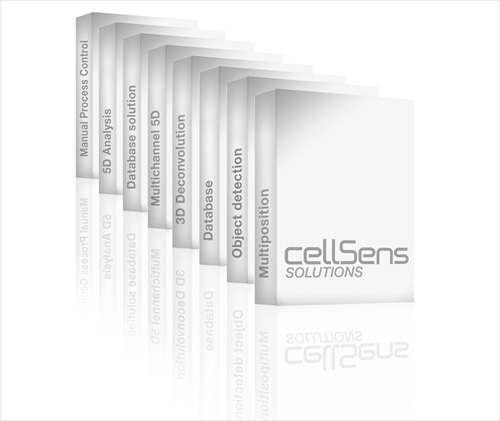
Olympus enhances its core cellSens life science imaging and analysis software with the release of version 1.8. New customisable interface and improved support for multi well plates enhance the user experience.
Focusing on ease of use for routine applications, the latest cellSens software release, version 1.8, offers flexible and user-centric imaging, processing, analysing and reporting. New and enhanced functions include a fully customisable user interface, precise frame-by-frame sample navigation and easy one-button switching between wells. For a wide range of life science applications, cellSens provides easy control of the workflow. The software binds together Olympus frames, cameras and high-quality optics to form complete, highly precise and user-friendly microscopy systems....
SCiLS Lab is the software developed by SCiLS GmbH which allows the user-friendly statistical analysis of large MALDI imaging datasets
MALDI (Matrix-Assisted Laser Desorption Ionization) imaging is a mass spectrometric imaging technique that allows the untargeted measurement of proteins, peptides, lipids, drugs and metabolites directly from tissue. MALDI imaging has been successfully applied in various research fields. A particular area of application is the histopathology research concerned with the definition of molecular phenotypes in cancer, like expression patterns that are associated with progression of cancer, or the discovery of novel proteins associated with metastasis....
At Forum Labo & Biotech, Malvern Instruments will exhibit the latest materials and biophysical characterization technology and instrumentation, including Archimedes, the new Zetasizer Nano ZSP and an extended range of sample dispersion accessories for the Mastersizer 3000.
In addition to participating in the exposition, the company is also running a half-day user training course on particle size measurement by laser diffraction and size of nanoparticles by dynamic light scattering, which is free of charge to Forum Labo attendees....
New Product Portfolio Release Significantly Reduces Hands-on Time for Tissue-based Research
Definiens, the global leader of image and data analysis solutions for quantitative digital pathology, today announced at the 4th International Definiens Symposium on Harvard Medical School campus the launch of new software that improves productivity in tissue-based research and biomarker development. The new software extends the functionality of Definiens’ existing product portfolio and provides a significant quality control advantage, with up to a two-fold reduction in hands on time for researchers.
Detection efficiency of Andor iXon 885 low-light EMCCD camera captures fluorescent interference patterns built up by individual massive molecules at double slit grating
Described as the "most beautiful experiment in physics", Richard Feynman spoke of the double-slit experiment as 'the heart of quantum physics', emphasizing how the diffraction of individual particles at a grating is an unambiguous demonstration of wave-particle duality and contrary to classical physics. Matter-wave interference has been observed for almost 90 years, first for electrons, neutrons, atoms, and small molecules. The first single-electron/double-slit experiment to back-up Feynman's assertion that an individual electron itself can behave like a wave was reported by Tonomura's team in 1989....
Read MoreJPK reports on the applied research of Ioan Notingher at the University of Nottingham using AFM and the Tip Assisted Optics module to study individual nanotubes and fibrils.May 9, 2013
JPK Instruments, a world-leading manufacturer of nanoanalytic instrumentation for research in life sciences and soft matter, reports on the research studies of Dr Ioan Notingher, associate professor in the faculty of science at the University of Nottingham.
With a research interest in the development of new optical and spectroscopic methods for studying biomaterials, he is applying JPK's atomic force microscope (AFM) system, the NanoWizard®, and tip assisted optics module (TAO). The research programmes of the Nottingham Nanoscience group span a range of exciting and topical themes in state-of-the-art nanoscale science.
FLIR Systems new Application Story Book brings together a wealth of useful information for organisations using or considering using thermal imaging for a scientific or research and development application.
Available as a pdf download or a 44-page hard copy brochure - the Applications Story Book provides a useful introduction to thermal imaging, how thermal imaging cameras work and reasons why you might choose to use this technique. The following pages detail the large number of Science / R&D applications where thermal imaging has become a preferred technique because of the information it can provide, its ease-of-use and non-destructive / non-contact testing methodology....
Read MoreMethod development to estimate the purity of vesicle preparations using Nanoparticle Tracking Analysis at the Cardiff University School of MedicineMay 7, 2013
NanoSight reports on how Nanoparticle Tracking Analysis, NTA, is being used in the development of a method to estimate the purity of vesicle preparations by comparing the ratio of nano-vesicle counts to protein concentration. This work is reported by Dr Aled Clayton of the School of Medicine at Cardiff University in an original research article published in the Journal of Extracellular Vesicles.
The School of Medicine at Cardiff University is one of the largest medical schools in the United Kingdom, home to over 3000 students and staff. It is a major international center for teaching and research providing a vibrant community of medical endeavor. The School is based at the University Hospital of Wales in Cardiff, but is also embedded at other hospital sites in Cardiff including Velindre Cancer Centre...
Read MoreAgar provides improved standards for AFM with Geller NIST and NPL-traceable calibration samplesMay 7, 2013
Agar Scientific, a leading supplier of microscopy accessories and consumables, provides NIST and NPL traceable Certified Reference Materials from Geller Microanalytical Laboratory.
As a leading supplier of accessories for microscopy, Agar Scientific has long been known for the quality of its wide range of calibration standards for Electron Microscopy. For AFM users, the Geller reference standards MRS-3, 4 and 5 are a series of high quality standards that are NIST/NPL traceable. They allow the accurate calibration of scanning probe instruments such as STM and AFM...
Read MoreLeica Microsystems and Leica Biosystems strengthen market position in BrazilMay 3, 2013
Long-Standing Leica Distributors Aotec Have Been Acquired
Leica Microsystems and Leica Biosystems have cemented ties with their Brazilian Leica distributor: All shares of Aotec Instrumentos Cientificos Ltda of São Paulo, Brazil, have been acquired. Aotec is a provider of microscopy and histopathology solutions and has been a Leica distributor for more than 25 years. All Aotec associates will remain with Aotec. Furthermore, the company is actively hiring additional staff to enlarge the Brazilian teams. Leica Microsystems and Leica Biosystems thereby strengthen their market position...
Read MoreMolecular Devices to exhibit at LaboTec Suisse 2013May 3, 2013
Molecular Devices will be showcasing their SpectraMax® i3 multi-mode microplate reader and AquaMax® Washer during LaboTec Suisse
The SpectraMax® i3 Multi-Mode Detection Platform is available as a stand-alone reader with three broad detection modes: luminescence, absorbance, and fluorescence. Users are then given the option to upgrade to additional applications and detection modes which expands the system's detection capability making it highly versatile, and able to offer application options far exceeding those of standard readers...
Unique QC and training software to help standardise blood film review
Haematology specialists, HORIBA UK Ltd – Medical, has introduced a new innovative service, The Quality Slide Program (QSP). Launched in France in 2012 to meet ISO guidelines for laboratory accreditation in blood film review and already used by over 40 laboratories, this program is now available in the UK. QSP is a digital blood cell morphology QC and training program. With the software, 6 digital blood films are published for download each month


 The all new Nikon SMZ25 and SMZ18 stereomicroscopes have evolved to meet the increasing needs for imaging systems that range from single cells to whole organisms. Featuring a large zoom ratio of 25:1 for the SMZ25, superior resolution and exceptional fluorescence transmission capability, the ergonomic, easy-to-use, fully motorised SMZ25 and manual SMZ18 are ideal for all bioscience applications....
 The Nano-mechanics group develops and uses novel techniques to understand molecular processes and has recently developed new instrumentation in the fields of single molecule force spectroscopy and correlation spectroscopy. There are three main areas of research: protein folding using AFM, measuring the mechanical properties of supported lipid bilayers and following the mechanical response of nano-confined fluids. This last area is performed on home-built instruments....
 Liquid-crystalline polymers (LCPs) are materials with exceptionally useful mechanical properties compared with ordinary industrial polymers. LCPs are not frequently used in industry due to their prohibitive production costs, but blending an LCP with a relatively common polymer is one way to utilize them at a reduced cost. Only blends that fibrillate in situ during processing, and when the fibrils maintain a solid state, do they demonstrate these superior properties...
 Scientists in the Laboratory of Microbiology at Wageningen UR are using a G:BOX Chemi system to accurately image both chemiluminescent and fluorescently labelled Western blots of multi-functional protein complexes belonging to the CRISPR/Cas proteins. These proteins degrade foreign DNA, thus protecting bacteria from infection and by studying them, the scientists hope to unravel how bacterial immune systems function. Determining pathways of bacterial immunity could lead to a number of applications, such as being able to immunise industrially important bacteria used in food production and large-scale fermentations against phage infection....
 At the 2013 Control international trade fair for quality assurance, ZEISS presented its new Hardware Auto Focus and cleanroom kit for the proven Axio Imager Vario microscope system. An integratable autofocus system ensures fast and precise focusing. The Hardware Auto Focus is ideal for surface inspections on reflective, low-contrast samples. In transmitted and reflected light (brightfield, darkfield, polarization contrast and oblique illumination), the focus system ensures high precision down to 0.3 of the depth of field of the objective lens....
 The TM3030 features improved electron optics to give better resolution and higher magnification capabilities together with built-in image processing to further enhance image quality. The improved electron-optical system of the TM3030 provides significantly better resolution, which is especially useful for the 5 kV mode of operation. Imaging at 5 kV reveals more surface detail and can be used for both topographic and elemental composition imaging. The new electron optics produce sharper images at higher magnifications across its three imaging and analysis modes....
 This unique patent pending design provides rich phenotypic cellular information with well-based quantitative data. Equipped with BioTek’s patented Hybrid Technology™ for microplate detection, Cytation3 includes both high sensitivity filter-based detection and a flexible monochromator based system for unmatched versatility and performance. The upgradable fluorescence cellular imaging module provides researchers rich cellular visualization analysis without the complexity and expense of standard automated microscopes, making cell imaging more accessible to a larger number of research laboratories at an affordable price...
 In a recently published paper, a team from Saarland University's Biophysics and Cryotechnology department in Germany has demonstrated a powerful, Raman-scattering technique for monitoring and investigating cryopreserved samples. In particular, they confirmed unambiguously the detection of ice crystals in vitreous samples in situ at temperatures below -120°C....
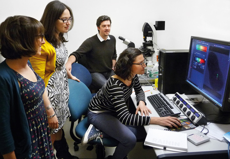 The Department of Biology at New York University has been equipped with the 100th Leica TCS SP8 X, a state-of-the-art confocal laser scanning microscope with a white light laser. The system offers complete spectral freedom for fluorescence experiments. For live cell imaging, the combination of high sensitivity and low laser power provides better cell viability. The new instrument is installed in the Center for Developmental Genetics and has been used to further Assistant Professor Dr. Lionel Christiaen’s goal of understanding how tissue-specific gene regulatory networks control and coordinate cell behavior during morphogenesis since February 2013....
 Focusing on ease of use for routine applications, the latest cellSens software release, version 1.8, offers flexible and user-centric imaging, processing, analysing and reporting. New and enhanced functions include a fully customisable user interface, precise frame-by-frame sample navigation and easy one-button switching between wells. For a wide range of life science applications, cellSens provides easy control of the workflow. The software binds together Olympus frames, cameras and high-quality optics to form complete, highly precise and user-friendly microscopy systems....
 MALDI (Matrix-Assisted Laser Desorption Ionization) imaging is a mass spectrometric imaging technique that allows the untargeted measurement of proteins, peptides, lipids, drugs and metabolites directly from tissue. MALDI imaging has been successfully applied in various research fields. A particular area of application is the histopathology research concerned with the definition of molecular phenotypes in cancer, like expression patterns that are associated with progression of cancer, or the discovery of novel proteins associated with metastasis....
 Definiens, the global leader of image and data analysis solutions for quantitative digital pathology, today announced at the 4th International Definiens Symposium on Harvard Medical School campus the launch of new software that improves productivity in tissue-based research and biomarker development. The new software extends the functionality of Definiens’ existing product portfolio and provides a significant quality control advantage, with up to a two-fold reduction in hands on time for researchers.
 and 2 min (b), 20 min (c), 40 min (d) and 90 min (e) after deposition. Scale bars, 20 micrometers (a–e). The colour bar ranges from 25 to 120 photons in a–d and from 220 to 650 photons in e. The arrow pointing downwards indicates the direction of the gravitational acceleration g.") Described as the "most beautiful experiment in physics", Richard Feynman spoke of the double-slit experiment as 'the heart of quantum physics', emphasizing how the diffraction of individual particles at a grating is an unambiguous demonstration of wave-particle duality and contrary to classical physics. Matter-wave interference has been observed for almost 90 years, first for electrons, neutrons, atoms, and small molecules. The first single-electron/double-slit experiment to back-up Feynman's assertion that an individual electron itself can behave like a wave was reported by Tonomura's team in 1989....
Described as the "most beautiful experiment in physics", Richard Feynman spoke of the double-slit experiment as 'the heart of quantum physics', emphasizing how the diffraction of individual particles at a grating is an unambiguous demonstration of wave-particle duality and contrary to classical physics. Matter-wave interference has been observed for almost 90 years, first for electrons, neutrons, atoms, and small molecules. The first single-electron/double-slit experiment to back-up Feynman's assertion that an individual electron itself can behave like a wave was reported by Tonomura's team in 1989....
 With a research interest in the development of new optical and spectroscopic methods for studying biomaterials, he is applying JPK's atomic force microscope (AFM) system, the NanoWizard®, and tip assisted optics module (TAO). The research programmes of the Nottingham Nanoscience group span a range of exciting and topical themes in state-of-the-art nanoscale science.
 Available as a pdf download or a 44-page hard copy brochure - the Applications Story Book provides a useful introduction to thermal imaging, how thermal imaging cameras work and reasons why you might choose to use this technique. The following pages detail the large number of Science / R&D applications where thermal imaging has become a preferred technique because of the information it can provide, its ease-of-use and non-destructive / non-contact testing methodology....
 with members of his group, Dr Joanne Welton, Dr Jason Webber & Miss Ridwana Chowdhury, with their NanoSight NTA system") The School of Medicine at Cardiff University is one of the largest medical schools in the United Kingdom, home to over 3000 students and staff. It is a major international center for teaching and research providing a vibrant community of medical endeavor. The School is based at the University Hospital of Wales in Cardiff, but is also embedded at other hospital sites in Cardiff including Velindre Cancer Centre...
The School of Medicine at Cardiff University is one of the largest medical schools in the United Kingdom, home to over 3000 students and staff. It is a major international center for teaching and research providing a vibrant community of medical endeavor. The School is based at the University Hospital of Wales in Cardiff, but is also embedded at other hospital sites in Cardiff including Velindre Cancer Centre...
 As a leading supplier of accessories for microscopy, Agar Scientific has long been known for the quality of its wide range of calibration standards for Electron Microscopy. For AFM users, the Geller reference standards MRS-3, 4 and 5 are a series of high quality standards that are NIST/NPL traceable. They allow the accurate calibration of scanning probe instruments such as STM and AFM...
 Leica Microsystems and Leica Biosystems have cemented ties with their Brazilian Leica distributor: All shares of Aotec Instrumentos Cientificos Ltda of São Paulo, Brazil, have been acquired. Aotec is a provider of microscopy and histopathology solutions and has been a Leica distributor for more than 25 years. All Aotec associates will remain with Aotec. Furthermore, the company is actively hiring additional staff to enlarge the Brazilian teams. Leica Microsystems and Leica Biosystems thereby strengthen their market position...
 The SpectraMax® i3 Multi-Mode Detection Platform is available as a stand-alone reader with three broad detection modes: luminescence, absorbance, and fluorescence. Users are then given the option to upgrade to additional applications and detection modes which expands the system's detection capability making it highly versatile, and able to offer application options far exceeding those of standard readers...
 Haematology specialists, HORIBA UK Ltd – Medical, has introduced a new innovative service, The Quality Slide Program (QSP). Launched in France in 2012 to meet ISO guidelines for laboratory accreditation in blood film review and already used by over 40 laboratories, this program is now available in the UK. QSP is a digital blood cell morphology QC and training program. With the software, 6 digital blood films are published for download each month