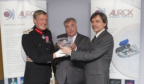
Oxfordshire company receives recognition for making confocal microscopy “personal”
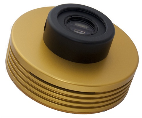
Aurox Limited, a leader in the design and manufacture of innovative optical imaging equipment, has won a 2012 Queen’s Award for Enterprise in Innovation. The award comes in recognition of the development and successful market introduction of the SD62 microscope, one of an ever increasing portfolio of Aurox products...
Read MoreArtemis CCD Launches QS14, Low-Light Imaging CCD CameraOct 9, 2012
For Real-time Results in RAMAN Spectroscopy, Chemiluminescence and Microscopy Applications
Norwich, UK: Artemis CCD, a leading manufacturer of cooled CCD cameras for low-light scientific imaging applications, is delighted to introduce its new QS14 thermoelectrically cooled CCD camera. This versatile camera is ideal for OEM manufacturers and scientists that need rapid, high quality images in low light conditions...
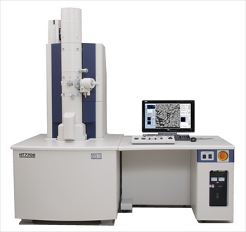
Read MoreHitachi's advanced HT7700 120kV TEM becomes more powerful!Oct 8, 2012
The introduction of the new objective lens option and a new STEM version makes Hitachi’s HT7700 the most powerful 120 kV TEM currently available, as well as the most intuitive
Now the ground-breaking HT7700 is just as well suited to the low accelerating voltage, high resolution examination of engineered light element materials as the advanced biomedical and biological samples for which it is already known. The new high resolution pole piece option for the HT7700 utilises Hitachi's unique double-gap objective lens technology, together with minimised spherical aberration to provide enhanced resolution – the best in class for a 120 kV instrument...
Read MoreIncrease Diagnostic Confidence and Laboratory Productivity with new Novocastra HD Antibodies for IHCOct 8, 2012
Leica Microsystems announces the introduction of the Novocastra™ HD antibody range, 80 clinically important immunohistochemistry antibody clones, spanning 10 anatomical pathologies
Market leaders in temperature controlled microscopy, Linkam Scientific Instruments report on the use of their FTIR600 stage at the University of Rome Tre to visualise and measure the in-situ dehydration of silicate materials
Geologists are interested in the distribution of volatile constituents across mineral crystals as this can provide insight into the crystallization process. This is now possible to investigate using modern FTIR imaging capabilities. On the Earth's crust, water bearing minerals are common. How these minerals dehydrate is important as this can give clues about dehydration-induced earthquakes and phase transformations at non-ambient conditions...
Read MoreSyngene's G:BOX XR5 is being utilised at the University of Cambridge to Contribute to Understanding which Genes are Involved in RemyelinationOct 8, 2012
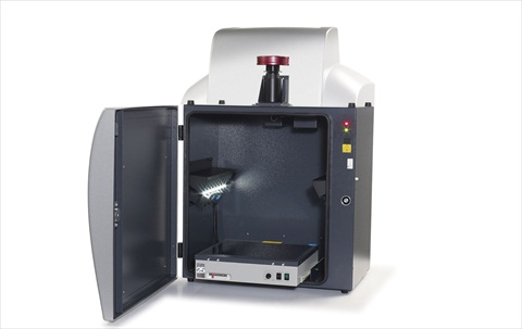
Syngene, a world-leading manufacturer of image analysis solutions is delighted to announce that the G:BOX XR5 imaging system is being used at the University of Cambridge to visualise and analyse DNA as part of a research programme to understand the molecular mechanisms behind why the remyelination process fails in diseases such as Multiple Sclerosis (MS)
Scientists in the Neurosciences Group at the Department of Veterinary Medicine, part of the University of Cambridge are using a G:BOX XR5 system to accurately image gels of fluorescent dye stained PCR products derived from genes involved in the remyelination process. By studying these genes, the scientists hope to find a means of enhancing the repair of normally non-repairable clinical conditions such as MS.
Read MoreRecord number of delegates and visitors flock to the 15th European Microscopy CongressOct 5, 2012
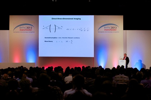
The 15th European Microscopy Congress – emc2012 - held at Manchester Central in September, was the largest yet in the series. It attracted 1,714 registered Conference Delegates. This is a 30% increase on 2008
The record breaking numbers were drawn by eight parallel conference sessions that embraced the life and physical sciences, and delivered a balanced programme of optical and electron microscopy. This breadth made emc2012 the most inclusive event yet. Professor Tony Wilson, President of the Royal Microscopical Society and Vice Chair of the Congress, said, “The conference sessions have provided an unparalleled opportunity for delegates to immerse themselves in their own area of interest, and also to witness new techniques and tools that might benefit their current work, or feature in their future activities.”...
Read MoreNon-invasive cytometric technique could eliminate need for blood samplesOct 3, 2012
Andor Newton camera powers novel non-invasive probe capable of providing cell concentration and morphological data for blood in vivo
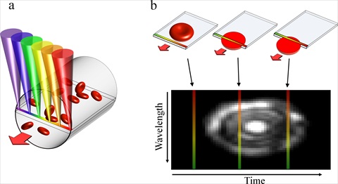
An Israeli team has demonstrated a non-invasive technique for imaging blood cells in vivo that could eliminate the need to extract blood from many patients. Powered by the Andor Newton Electron Multiplying EMCCD camera, their high-resolution Spectrally Encoded Flow Cytometry (SEFC) probe offers primary care physicians the capability to detect directly a wide range of common medical disorders, such as anaemia and bacterial infection, and potentially life threatening conditions, including sepsis, thrombosis and sickle cell crisis...
Read MoreCarl Zeiss introduces ORION NanoFabOct 3, 2012
Extending nanofabrication to the sub-10 nanometer scale
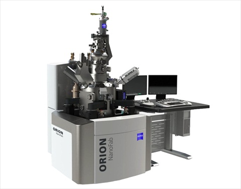
Carl Zeiss Microscopy has recently introduced ORION NanoFab at the European Microscopy Congress (EMC) in Manchester, UK. It is the first multi-ion-beam tool based on Gas Field Ion Source (GFIS) technology. As a major enhancement to the existing helium ion microscope, ORION NanoFab also utilizes neon ions. The system is therefore capable of providing a complete sub-10 nanometer nanofabrication and sub-nanometer imaging solution for industry, government, and academic research laboratories. An optional gallium focused ion beam (FIB) column can also be integrated...
Read MoreBenefit from even more advanced material science imaging and analysis with the LEXT OLS4000 software version 2.2Oct 3, 2012
Staying at the forefront of optical metrology
Setting a new standard of performance in measuring technology, Olympus has released an updated version of its LEXT OLS4000 confocal laser scanning microscope (CLSM) software. Compatible with Windows 7, 64 bit operation, the LEXT software version 2.2 allows users to capture high resolution images up to 10 times faster than the previous version. In addition, the new 3D multilayer function can measure transparent layers, allowing the accurate measurement and subsequent analysis of multiple layers within a single sample...
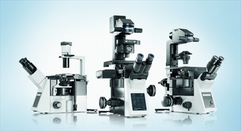
Read MoreThe next generation of inverted microscope systems has arrivedSep 26, 2012
The new Olympus IX3 series with intuitive ease-of-use and flexible ‘open access’ light path
Olympus has released the innovative new IX3 series of inverted research microscope systems for effortless, intuitive live cell imaging and clinical analysis. This includes the fully automated IX83 for high-end research applications, the flexible IX73, which can be configured in manual, semi-motorised or motorised modes, and the easy-to-use IX53 with fluorescent capabilities, which is optimised for the routine examination of tissue samples. Built using worldwide customer feedback and designed to meet the needs of a wide range of users, the new systems offer exceptional ease-of-use and unprecedented optical flexibility via a new, customisable light path...
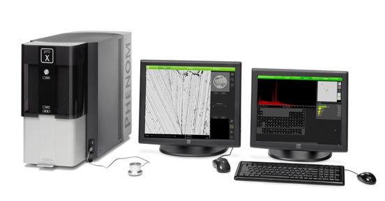
Read MoreFast and smart study of environmental pollution with desktop SEM Phenom proXSep 24, 2012
The importance of environmental quality demands that biologists both understand natural ecosystems and learn to be effective problem solvers
One key environmental issue involves the effects of the pollution from heavy industries on the environment. Different equipment with different techniques is required to be able to study and analyze these effects. With the Phenom proX desktop SEM, sample structures can be physically examined and their elemental composition determined. Viewing three-dimensional images of microscopic structures only solves half the problem when analyzing samples. It is often necessary to collect more than optical data to be able to identify the different elements in a specimen...
Read MoreQueensgate to launch Dual Sensor Technology at emc2012Sep 10, 2012
Queensgate Instruments - an Elektron Technology brand - is to unveil its revolutionary new Dual Sensor Technology at this year's European Microscopy Congress (emc2012) from the 16th to 21st of September in Manchester
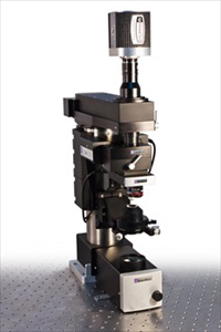
JPK Instruments, a world-leading manufacturer of nanoanalytic instrumentation for research in life sciences and soft matter, reports on the use of AFM systems in the group of Dr Bart Hoogenboom of the London Centre for Nanotechnology
Lecturer at the London Nanotechnology Centre and the Department of Physics and Astronomy, University College London, Dr Bart Hoogenboom's main research interest is where nanotechnology tools may be used to study and manipulate single biomolecules. Specifically, he applies AFM as the only instrument that provides ~1 nm spatial resolution on large biomolecules that are "alive", or in more scientific terms, they are still functional and may be studied in the natural environment, i.e. aqueous salt solution...
Read MoreAgar Scientific reports on the MAC range of reference and calibration standards available for microanalysis usersSep 4, 2012
Agar Scientific, a leading supplier of microscopy accessories and consumables, reports on the range of reference and calibration standards available for scanning electron microscopes manufactured by Micro-Analysis Consultants
Agar Scientific is a market leader in the supply of high quality accessories to assist with sample handling for the microscopy market serving a very broad range of application areas. With traceability of results becoming ever more important, the availability of reliable calibration standards is extremely important to the electron microscopist...
Read MoreGround-breaking versatility from ScientificaSep 3, 2012
Multiphoton imaging is one of the most powerful life science research tools of the 21st century and Scientifica has acknowledged this with the launch of a modular Multiphoton Imaging System
For the first time, it allows the user to build a cost effective and future proof system based around the renowned SliceScope microscope. Multiphoton imaging uses high intensity pulsed lasers, fast moving mirrors, and precision optics to allow researchers to image structure and activity within living tissue. A laser spot is scanned quickly over a sample, exciting dyes within the tissue. The emitted light is collected with sensitive photomultiplier tubes (PMTs) and the image is captured and displayed on a PC screen...
Read MoreGuaranteed Knockdown for siRNA Gene SilencingSep 3, 2012
AMSBIO has announced its new Trilencer-27 siRNA kit, that contains Dicer-Substrate duplexes, provides critical improvements over the use of traditional 21mer siRNA designs
Gene silencing through the use of RNAi has become a primary tool for characterizing gene involvement in disease states and interactive pathways. Offering genome-wide coverage against human, mouse and rat, the AMSBIO Trilencer-27 siRNA kit takes advantage of the natural processing by Dicer to produce 10-fold higher potency and specificity than shorter 21mer RNAi forms. Beneficially the 27mer dicer-substrate duplexes in the kit also evade the radar of the mammalian interferon response when expressed in mammalian cells and initiate strong and specific gene silencing...
Read MoreMacro to Micro Fluorescence In Vivo Imaging with the iBox Explorer2 Imaging MicroscopeAug 30, 2012
The iBox® Explorer™2 Imaging Microscope now features expanded capabilities for detection of fluorescent markers in small animals
With magnification from whole mouse to individual cells, the iBox Explorer2 provides breakthrough advances for cancer research studies. Researchers can now visualize tumor margins, micro metastasis and more with the wide range of magnifications from 0.17x to 16.5x. The upright optics provide an ultra-long working distance and high numerical aperture (NA) for detailed, exacting in vivo imaging research...
University of Leicester scientists develop rapid-scanning microscope with no loss of quality
Researchers at the University of Leicester have developed a new form of digital microscope which can create an image 100 times faster than regular equipment - without losing image quality. The team of scientists have developed a new type of confocal microscope that produces high-resolution images at very fast speeds...
Read MoreCarl Zeiss wins G.I.T. InnovationsAward 2012 for AURIGA LaserAug 29, 2012
The G.I.T. Laboratory Journal officially announced the winners of the G.I.T. InnovationsAward 2012. The CrossBeam (FIB SEM) AURIGA Laser from Carl Zeiss Microscopy took first place in the field of Laboratory Equipment & Technology
Prior to the final voting, a jury of scientists had examined all submissions and made a selection. Readers of G.I.T. Laboratory Journal, customers and visitors of Analytica 2012 in Munich and of Achema 2012 in Frankfurt then selected their favorites. Carl Zeiss Microscopy launched the FIB SEM in March this year. The system combines the advantages of the CrossBeam workstation with the capabilities of a pulsed micro-focus laser for fast ablation of material...
Read MoreAutomating Cell-Based Microplate AssaysAug 27, 2012
PHERAstar FS with Direct Optic Bottom Reading, the Optimal Solution for Cell Based Assays
One of the most important uses for fluorescent proteins like GFP, mCherry, and mOranage is real-time monitoring of cellular processes in live cells. Now with the PHERAstar FS microplate reader, it is possible to miniaturize and increase throughput of fluorescent protein cell-based assays that were not possible before...
Market leaders in temperature controlled microscopy, Linkam Scientific Instruments, have been chosen as suppliers of a CSS450 for the Polymeric and Mesomorphic Materials Group, CENIMAT, Portugal for the rheological characterization of anisotropic materials
CENIMAT is a national scientific research center sponsored by the Portuguese Ministry of Science, Technology and Higher Education. It is divided into four Research Teams: Dielectrics Materials and Structures Group, Electronics and Microelectronics Materials Group, Polymeric and Mesomorphic Materials Group and Structural Materials Group.
Read More


 Aurox Limited, a leader in the design and manufacture of innovative optical imaging equipment, has won a 2012 Queen’s Award for Enterprise in Innovation. The award comes in recognition of the development and successful market introduction of the SD62 microscope, one of an ever increasing portfolio of Aurox products...
 Norwich, UK: Artemis CCD, a leading manufacturer of cooled CCD cameras for low-light scientific imaging applications, is delighted to introduce its new QS14 thermoelectrically cooled CCD camera. This versatile camera is ideal for OEM manufacturers and scientists that need rapid, high quality images in low light conditions...
 Now the ground-breaking HT7700 is just as well suited to the low accelerating voltage, high resolution examination of engineered light element materials as the advanced biomedical and biological samples for which it is already known. The new high resolution pole piece option for the HT7700 utilises Hitachi's unique double-gap objective lens technology, together with minimised spherical aberration to provide enhanced resolution – the best in class for a 120 kV instrument...
 Novocastra HD was developed to meet the needs of pathologists and histologists, in terms of menu, diagnostic confidence and workflow. This new ‘Highly Definitive’ range was created following extensive global market research on customer preferences, combined with the results of a comprehensive competitive evaluation* of each antibody by NordiQC. The Novocastra HD menu therefore represents antibodies with independently qualified performance, each available in a range of new sizes and formats aligned to laboratory workloads...
 Geologists are interested in the distribution of volatile constituents across mineral crystals as this can provide insight into the crystallization process. This is now possible to investigate using modern FTIR imaging capabilities. On the Earth's crust, water bearing minerals are common. How these minerals dehydrate is important as this can give clues about dehydration-induced earthquakes and phase transformations at non-ambient conditions...
 Scientists in the Neurosciences Group at the Department of Veterinary Medicine, part of the University of Cambridge are using a G:BOX XR5 system to accurately image gels of fluorescent dye stained PCR products derived from genes involved in the remyelination process. By studying these genes, the scientists hope to find a means of enhancing the repair of normally non-repairable clinical conditions such as MS.
 The record breaking numbers were drawn by eight parallel conference sessions that embraced the life and physical sciences, and delivered a balanced programme of optical and electron microscopy. This breadth made emc2012 the most inclusive event yet. Professor Tony Wilson, President of the Royal Microscopical Society and Vice Chair of the Congress, said, “The conference sessions have provided an unparalleled opportunity for delegates to immerse themselves in their own area of interest, and also to witness new techniques and tools that might benefit their current work, or feature in their future activities.”...
 An Israeli team has demonstrated a non-invasive technique for imaging blood cells in vivo that could eliminate the need to extract blood from many patients. Powered by the Andor Newton Electron Multiplying EMCCD camera, their high-resolution Spectrally Encoded Flow Cytometry (SEFC) probe offers primary care physicians the capability to detect directly a wide range of common medical disorders, such as anaemia and bacterial infection, and potentially life threatening conditions, including sepsis, thrombosis and sickle cell crisis...
 Carl Zeiss Microscopy has recently introduced ORION NanoFab at the European Microscopy Congress (EMC) in Manchester, UK. It is the first multi-ion-beam tool based on Gas Field Ion Source (GFIS) technology. As a major enhancement to the existing helium ion microscope, ORION NanoFab also utilizes neon ions. The system is therefore capable of providing a complete sub-10 nanometer nanofabrication and sub-nanometer imaging solution for industry, government, and academic research laboratories. An optional gallium focused ion beam (FIB) column can also be integrated...
 Olympus has released the innovative new IX3 series of inverted research microscope systems for effortless, intuitive live cell imaging and clinical analysis. This includes the fully automated IX83 for high-end research applications, the flexible IX73, which can be configured in manual, semi-motorised or motorised modes, and the easy-to-use IX53 with fluorescent capabilities, which is optimised for the routine examination of tissue samples. Built using worldwide customer feedback and designed to meet the needs of a wide range of users, the new systems offer exceptional ease-of-use and unprecedented optical flexibility via a new, customisable light path...
 One key environmental issue involves the effects of the pollution from heavy industries on the environment. Different equipment with different techniques is required to be able to study and analyze these effects. With the Phenom proX desktop SEM, sample structures can be physically examined and their elemental composition determined. Viewing three-dimensional images of microscopic structures only solves half the problem when analyzing samples. It is often necessary to collect more than optical data to be able to identify the different elements in a specimen...
 This event is home to Europe's largest exhibition dedicated to microscopy, and will see the launch of Queensgate's groundbreaking new objective scanning mechanism and analogue controller, offering faster, more accurate and more stable microscope objective focusing than ever before...
 Lecturer at the London Nanotechnology Centre and the Department of Physics and Astronomy, University College London, Dr Bart Hoogenboom's main research interest is where nanotechnology tools may be used to study and manipulate single biomolecules. Specifically, he applies AFM as the only instrument that provides ~1 nm spatial resolution on large biomolecules that are "alive", or in more scientific terms, they are still functional and may be studied in the natural environment, i.e. aqueous salt solution...
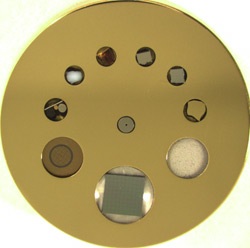 Agar Scientific is a market leader in the supply of high quality accessories to assist with sample handling for the microscopy market serving a very broad range of application areas. With traceability of results becoming ever more important, the availability of reliable calibration standards is extremely important to the electron microscopist...
 For the first time, it allows the user to build a cost effective and future proof system based around the renowned SliceScope microscope. Multiphoton imaging uses high intensity pulsed lasers, fast moving mirrors, and precision optics to allow researchers to image structure and activity within living tissue. A laser spot is scanned quickly over a sample, exciting dyes within the tissue. The emitted light is collected with sensitive photomultiplier tubes (PMTs) and the image is captured and displayed on a PC screen...

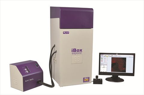 With magnification from whole mouse to individual cells, the iBox Explorer2 provides breakthrough advances for cancer research studies. Researchers can now visualize tumor margins, micro metastasis and more with the wide range of magnifications from 0.17x to 16.5x. The upright optics provide an ultra-long working distance and high numerical aperture (NA) for detailed, exacting in vivo imaging research...
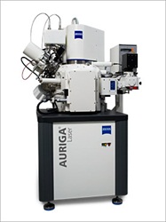 Prior to the final voting, a jury of scientists had examined all submissions and made a selection. Readers of G.I.T. Laboratory Journal, customers and visitors of Analytica 2012 in Munich and of Achema 2012 in Frankfurt then selected their favorites. Carl Zeiss Microscopy launched the FIB SEM in March this year. The system combines the advantages of the CrossBeam workstation with the capabilities of a pulsed micro-focus laser for fast ablation of material...
 One of the most important uses for fluorescent proteins like GFP, mCherry, and mOranage is real-time monitoring of cellular processes in live cells. Now with the PHERAstar FS microplate reader, it is possible to miniaturize and increase throughput of fluorescent protein cell-based assays that were not possible before...
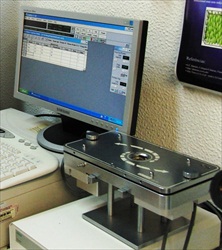 CENIMAT is a national scientific research center sponsored by the Portuguese Ministry of Science, Technology and Higher Education. It is divided into four Research Teams: Dielectrics Materials and Structures Group, Electronics and Microelectronics Materials Group, Polymeric and Mesomorphic Materials Group and Structural Materials Group.