PerkinElmer, Inc., a global leader committed to innovating for a healthier world, has launched the first cell painting kit as part of its new portfolio of PhenoVue™ cellular imaging reagents. This new range of reagents leverages PerkinElmer’s expertise in cellular imaging and high-content screening and works alongside the Company’s microplates, automation, software and industry-leading high-content screening instruments....
Linkam Scientific’s PE120 Peltier system formed part of the experimental set-up used to develop a radical new diagnostic test for the SARS CoV-2 virus at Ohio State University (OSU), Columbus, Ohio, USA. The global outbreak of COVID-19 has prompted scientists to focus their research on rapid and robust diagnostic tests. Reverse-transcription polymerase chain reaction (RT-PCR) assays – the ‘gold-standard’ for molecular clinical diagnostics – became available quickly, however this provides relatively long characterisation times...
To take AI development in pathology to the next level, a European consortium combining leading European research centres, hospitals as well as major pharmaceutical industries, is going to develop a repository for the sharing of pathology data. The 6-year, €70 million project called BIGPICTURE, will herald a new era in pathology. Sectra engages in the new EU Innovative Medicines Initiative project BIGPICTURE to construct a large-scale database of pathology images....
Now available from Alpha Laboratories, the new gelLITE gel documentation system is a compact and convenient system for documentation of agarose gels and stained protein gels. A 302nm UV transilluminator enables imaging of large format agarose gels or stained protein gels up to 20 x 20 cm, and can slide out from the main body of the system allowing gel extraction of DNA fragments...
Ahead of the upcoming missions to search for evidence of life on Mars, researchers used Linkam’s HFS600 stage in temperature-controlled experiments to study the formation of mixed sulfates – a key indicator of biological processes on the red planet. NASA’s Mars 2020 Perseverance rover will land on Mars on 18 February 2021, to search for signs of life and explore the planet's geology....
ZEISS has introduced a new compact microscope for digital teaching and routine lab work just in time for the International Day of Education, which took place yesterday. ZEISS Primostar 3 is a robust upright light microscope, which is made for daily work in a classroom or in a lab for tissue and sample examination in histology, cell biology, food or microbiology, etc. It is designed for long-term use and extreme durability....
Tomocube’s cutting-edge 3D quantitative phase imaging is set to play a crucial role in platelet research according to a new paper from scientists at the University of York. Using holotomography images generated by the Tomocube HT-2H microscope, the team were able to identify and quantify in single unlabelled, live platelets clear disparities in activation status and potential functional ability in disease states without experimental interference, such as from fixation or labelling...
Complementary metal-oxide-semiconductor (CMOS) technology now offers the advanced imaging capabilities required for many biomedical applications, but can it replace the more expensive sCMOS (scientific CMOS) sensors? CMOS and sCMOS sensors have set the benchmark for both performance and value in machine vision in several industries, and this article will explain the benefits and costs of each technology for highly demanding imaging applications in biomedical and life sciences....
Making people stand in front of a scanner to have their body temperature read can result in a large number of false negatives, allowing people with Covid-19 to pass through airports and hospitals undetected. Leading experts in physiology have suggested instead that taking temperature readings of a person’s fingertip and eye would give a significantly better and more reliable reading and help identify those with fever....
ZEISS introduces ZEISS Solutions Lab, an innovative service, that provides its customers with rapid access to new applications for their microscopy systems or correlative microscopy suites. ZEISS Solutions Lab enables customers to select, or to drive demand for module-based workflows built on ZEISS software packages for a wide variety of requirements specific to their research needs...
New research on the structure and dynamics of a branched form of DNA called a three-way junction could lead to more effectively targeted treatments for degenerative disorders like Huntington’s Disease, scientists say. In a new paper published in the journal Nature Communications, chemists from the University of Glasgow show for the first time how three-way DNA junctions undergo unexpected rearrangements in their structure....
ZEISS announces that it has formed a research collaboration partnership with the Max Planck Florida Institute for Neuroscience (MPFI). Using an LSM 980 NLO next generation confocal microscope supplied by ZEISS, MPFI will investigate using implanted GRadient INdex (GRIN) lenses in combination with the Airyscan 2 area detector for deep brain functional neuroscience research....
Upgrading a microscope with a motorized stage enhances a microscope’s capability by allowing for rapid, smooth, and highly repeatable sample movement. This can be difficult to achieve or impractical, using a manual stage, especially when the experiment requires the operator to perform repeated, precise, and accurate movements over a long time period. Motorized stages allow the user to pre-program movements, set and save location points for return inspection, and integrate the stage’s positioning within the imaging process....
JEOL USA awarded two Grand Prizes to winners of its 2020 Electron Microscopy Image Contest, and kicked off its 2021 Image Contest at the beginning of the new year. The annual contest showcases JEOL microscope users’ artistically or esthetically pleasing images with good composition, sharp focus, and technical competency, especially in the use of accelerating voltage...
ZEISS is expanding its imaging technology capabilities with 3D and big image data software by acquiring a majority stake in the imaging business of arivis. With this investment, ZEISS is further strengthening its software competencies and market position in 3D image visualization, image processing, and analysis software for research microscopy. Both companies have a long-standing collaboration and strategic partnership, as arivis has been an important development partner for ZEISS for more than seven years..
Merck announce the launch of its new Scepter™ 3.0 handheld automated cell counter, a solution that enables convenient, on-demand cell volume, cell diameter and cell concentration data. The device uses precision microfluidics to measure and count cells via electrical impedance within 30 seconds. The Scepter™ 3.0 cell counter offers researchers true automation without the risk of error that accompanies vision-based systems, using a combination of analog and digital hardware for sensing, signal processing, data storage, and graphical display....
With the release of ZEISS Lattice Lightsheet 7, ZEISS places a turn-key lattice light sheet instrument at the disposal of the life science research community. Based on the pioneering research and developments of Ernst H. K. Stelzer while at EMBL, Heidelberg, on light sheet technology and of Nobel laureate Eric Betzig while at the Janelia Research Campus of HHMI on structuring light sheets as optical lattices to render them thinner and longer...
Add powerful automated autofocus to your existing microscope. Prior Scientific’s PureFocus850 Laser Autofocus combines advanced optics and an on board microprocessor to provide real time focus control for infinity corrected optical systems. The PureFocus850 allows for rapid scanning of biological and industrial samples, dramatically reducing scan time and improving throughput....
ZEISS presents a new generation of its field emission scanning electron microscope (FESEM) family ZEISS GeminiSEM. The new models ZEISS GeminiSEM 360, 460 and 560 are tailored for sub-nanometer imaging and effortless analytics. Users benefit from innovations in the electron optics and a new chamber design offering better image quality, usability and flexibility. ZEISS GeminiSEM 560 now being introduced brings the ZEISS Gemini 3 column to the market for the first time....
Hologic, Inc., has announced that its new Genius™ Digital Diagnostics System is now CE marked in Europe. Genius Digital Diagnostics is the first digital cytology platform to combine a new artificial intelligence (AI) algorithm with advanced digital imaging to help cytotechnologists and pathologists identify pre-cancerous lesions and cancer cells in women....
The AutoCOL fully automated colony counting system allows the user to count colonies from 100 plates in 40 minutes, a task which no other colony counter currently on the market can perform.Built around Synbiosis’ gold standard ProtoCOL technology, AutoCOL can produce precise images of bacteria, yeast, and mould cultured on any agar plate. Compared to manual counting the AutoCOL has the capability to increase plate reading productivity by over 700 percent.
Edinburgh Instruments is delighted to announce the launch of the new RMS1000 Raman Microscope designed and manufactured at their global headquarters in Scotland. The RMS1000 Raman Microscope is an open architecture, research grade confocal Raman Microscope. It has been designed to be adapted to almost any modern, state-of-the-art Raman application. This high-end research tool has been built with no compromises; resulting in a system that stands alone in both specification and ease of use....

 PerkinElmer, Inc., a global leader committed to innovating for a healthier world, has launched the first cell painting kit as part of its new portfolio of PhenoVue™ cellular imaging reagents. This new range of reagents leverages PerkinElmer’s expertise in cellular imaging and high-content screening and works alongside the Company’s microplates, automation, software and industry-leading high-content screening instruments....
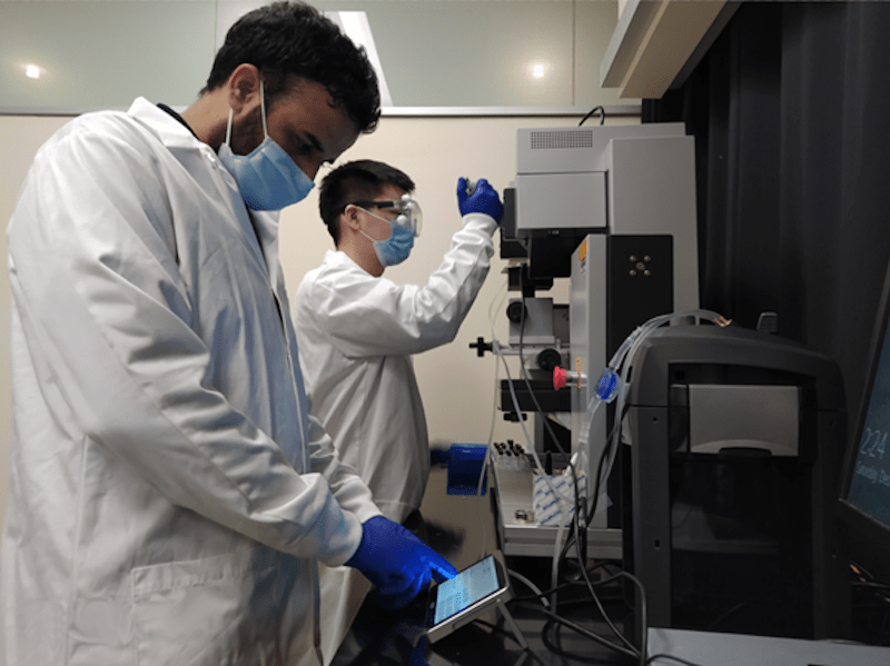 Linkam Scientific’s PE120 Peltier system formed part of the experimental set-up used to develop a radical new diagnostic test for the SARS CoV-2 virus at Ohio State University (OSU), Columbus, Ohio, USA. The global outbreak of COVID-19 has prompted scientists to focus their research on rapid and robust diagnostic tests. Reverse-transcription polymerase chain reaction (RT-PCR) assays – the ‘gold-standard’ for molecular clinical diagnostics – became available quickly, however this provides relatively long characterisation times...
 To take AI development in pathology to the next level, a European consortium combining leading European research centres, hospitals as well as major pharmaceutical industries, is going to develop a repository for the sharing of pathology data. The 6-year, €70 million project called BIGPICTURE, will herald a new era in pathology. Sectra engages in the new EU Innovative Medicines Initiative project BIGPICTURE to construct a large-scale database of pathology images....
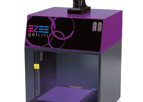 Now available from Alpha Laboratories, the new gelLITE gel documentation system is a compact and convenient system for documentation of agarose gels and stained protein gels. A 302nm UV transilluminator enables imaging of large format agarose gels or stained protein gels up to 20 x 20 cm, and can slide out from the main body of the system allowing gel extraction of DNA fragments...
 Ahead of the upcoming missions to search for evidence of life on Mars, researchers used Linkam’s HFS600 stage in temperature-controlled experiments to study the formation of mixed sulfates – a key indicator of biological processes on the red planet. NASA’s Mars 2020 Perseverance rover will land on Mars on 18 February 2021, to search for signs of life and explore the planet's geology....
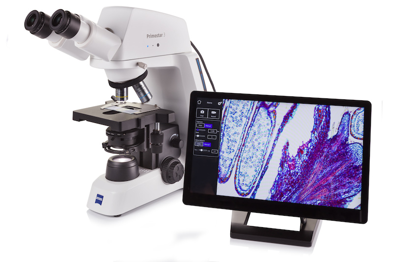 ZEISS has introduced a new compact microscope for digital teaching and routine lab work just in time for the International Day of Education, which took place yesterday. ZEISS Primostar 3 is a robust upright light microscope, which is made for daily work in a classroom or in a lab for tissue and sample examination in histology, cell biology, food or microbiology, etc. It is designed for long-term use and extreme durability....
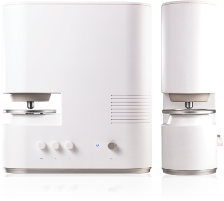 Tomocube’s cutting-edge 3D quantitative phase imaging is set to play a crucial role in platelet research according to a new paper from scientists at the University of York. Using holotomography images generated by the Tomocube HT-2H microscope, the team were able to identify and quantify in single unlabelled, live platelets clear disparities in activation status and potential functional ability in disease states without experimental interference, such as from fixation or labelling...
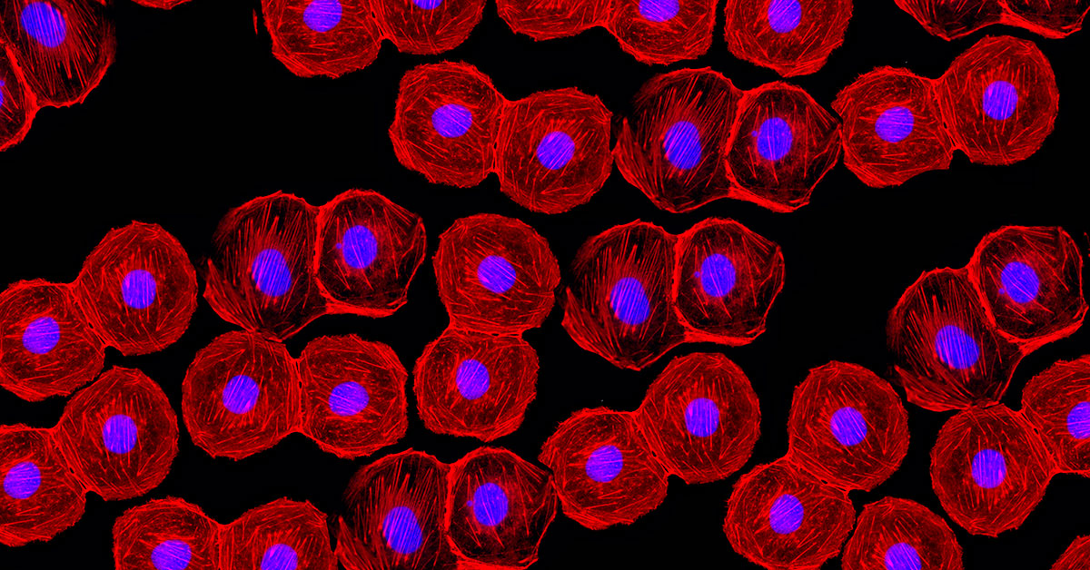 Complementary metal-oxide-semiconductor (CMOS) technology now offers the advanced imaging capabilities required for many biomedical applications, but can it replace the more expensive sCMOS (scientific CMOS) sensors? CMOS and sCMOS sensors have set the benchmark for both performance and value in machine vision in several industries, and this article will explain the benefits and costs of each technology for highly demanding imaging applications in biomedical and life sciences....
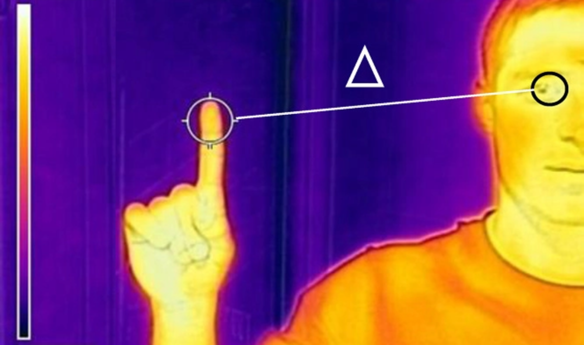 Making people stand in front of a scanner to have their body temperature read can result in a large number of false negatives, allowing people with Covid-19 to pass through airports and hospitals undetected. Leading experts in physiology have suggested instead that taking temperature readings of a person’s fingertip and eye would give a significantly better and more reliable reading and help identify those with fever....
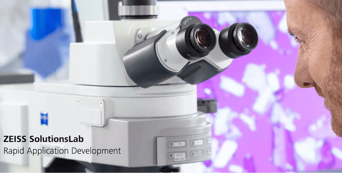 ZEISS introduces ZEISS Solutions Lab, an innovative service, that provides its customers with rapid access to new applications for their microscopy systems or correlative microscopy suites. ZEISS Solutions Lab enables customers to select, or to drive demand for module-based workflows built on ZEISS software packages for a wide variety of requirements specific to their research needs...
 New research on the structure and dynamics of a branched form of DNA called a three-way junction could lead to more effectively targeted treatments for degenerative disorders like Huntington’s Disease, scientists say. In a new paper published in the journal Nature Communications, chemists from the University of Glasgow show for the first time how three-way DNA junctions undergo unexpected rearrangements in their structure....
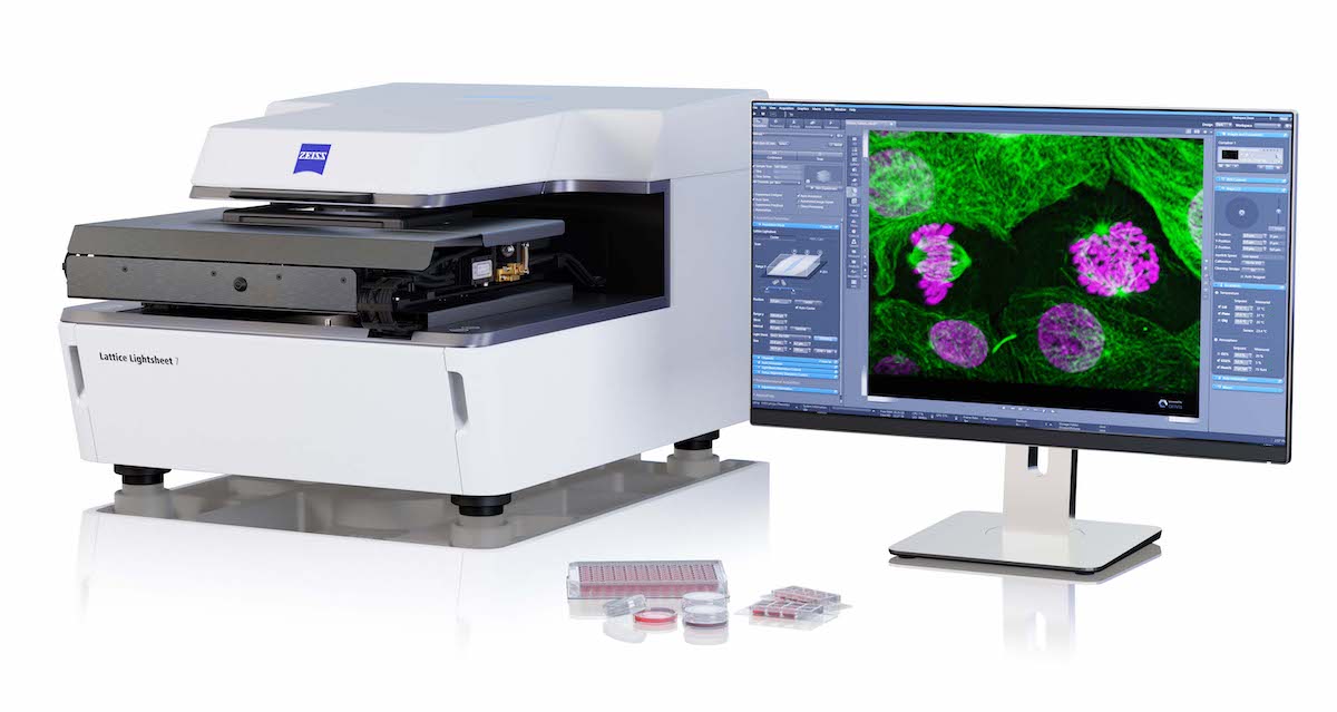 ZEISS announces that it has formed a research collaboration partnership with the Max Planck Florida Institute for Neuroscience (MPFI). Using an LSM 980 NLO next generation confocal microscope supplied by ZEISS, MPFI will investigate using implanted GRadient INdex (GRIN) lenses in combination with the Airyscan 2 area detector for deep brain functional neuroscience research....
 Upgrading a microscope with a motorized stage enhances a microscope’s capability by allowing for rapid, smooth, and highly repeatable sample movement. This can be difficult to achieve or impractical, using a manual stage, especially when the experiment requires the operator to perform repeated, precise, and accurate movements over a long time period. Motorized stages allow the user to pre-program movements, set and save location points for return inspection, and integrate the stage’s positioning within the imaging process....
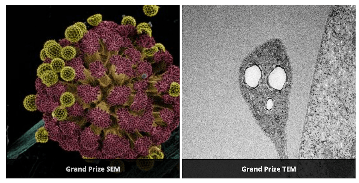 JEOL USA awarded two Grand Prizes to winners of its 2020 Electron Microscopy Image Contest, and kicked off its 2021 Image Contest at the beginning of the new year. The annual contest showcases JEOL microscope users’ artistically or esthetically pleasing images with good composition, sharp focus, and technical competency, especially in the use of accelerating voltage...
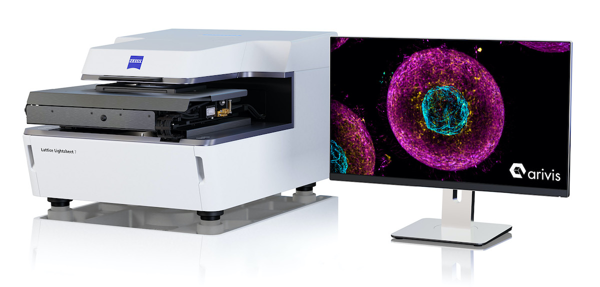 ZEISS is expanding its imaging technology capabilities with 3D and big image data software by acquiring a majority stake in the imaging business of arivis. With this investment, ZEISS is further strengthening its software competencies and market position in 3D image visualization, image processing, and analysis software for research microscopy. Both companies have a long-standing collaboration and strategic partnership, as arivis has been an important development partner for ZEISS for more than seven years..
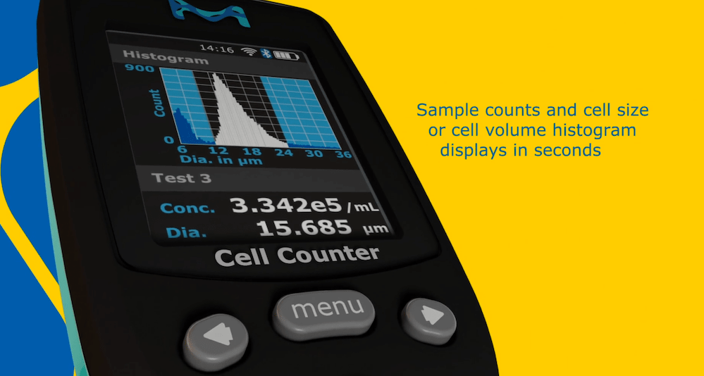 Merck announce the launch of its new Scepter™ 3.0 handheld automated cell counter, a solution that enables convenient, on-demand cell volume, cell diameter and cell concentration data. The device uses precision microfluidics to measure and count cells via electrical impedance within 30 seconds. The Scepter™ 3.0 cell counter offers researchers true automation without the risk of error that accompanies vision-based systems, using a combination of analog and digital hardware for sensing, signal processing, data storage, and graphical display....
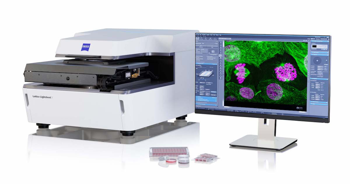 With the release of ZEISS Lattice Lightsheet 7, ZEISS places a turn-key lattice light sheet instrument at the disposal of the life science research community. Based on the pioneering research and developments of Ernst H. K. Stelzer while at EMBL, Heidelberg, on light sheet technology and of Nobel laureate Eric Betzig while at the Janelia Research Campus of HHMI on structuring light sheets as optical lattices to render them thinner and longer...
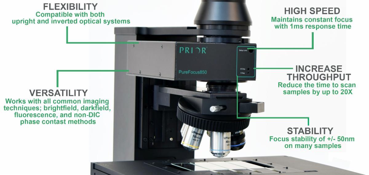 Add powerful automated autofocus to your existing microscope. Prior Scientific’s PureFocus850 Laser Autofocus combines advanced optics and an on board microprocessor to provide real time focus control for infinity corrected optical systems. The PureFocus850 allows for rapid scanning of biological and industrial samples, dramatically reducing scan time and improving throughput....
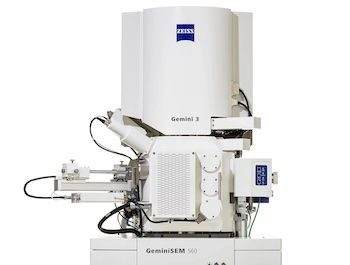 ZEISS presents a new generation of its field emission scanning electron microscope (FESEM) family ZEISS GeminiSEM. The new models ZEISS GeminiSEM 360, 460 and 560 are tailored for sub-nanometer imaging and effortless analytics. Users benefit from innovations in the electron optics and a new chamber design offering better image quality, usability and flexibility. ZEISS GeminiSEM 560 now being introduced brings the ZEISS Gemini 3 column to the market for the first time....
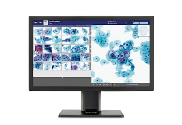 Hologic, Inc., has announced that its new Genius™ Digital Diagnostics System is now CE marked in Europe. Genius Digital Diagnostics is the first digital cytology platform to combine a new artificial intelligence (AI) algorithm with advanced digital imaging to help cytotechnologists and pathologists identify pre-cancerous lesions and cancer cells in women....
 The AutoCOL fully automated colony counting system allows the user to count colonies from 100 plates in 40 minutes, a task which no other colony counter currently on the market can perform.Built around Synbiosis’ gold standard ProtoCOL technology, AutoCOL can produce precise images of bacteria, yeast, and mould cultured on any agar plate. Compared to manual counting the AutoCOL has the capability to increase plate reading productivity by over 700 percent.
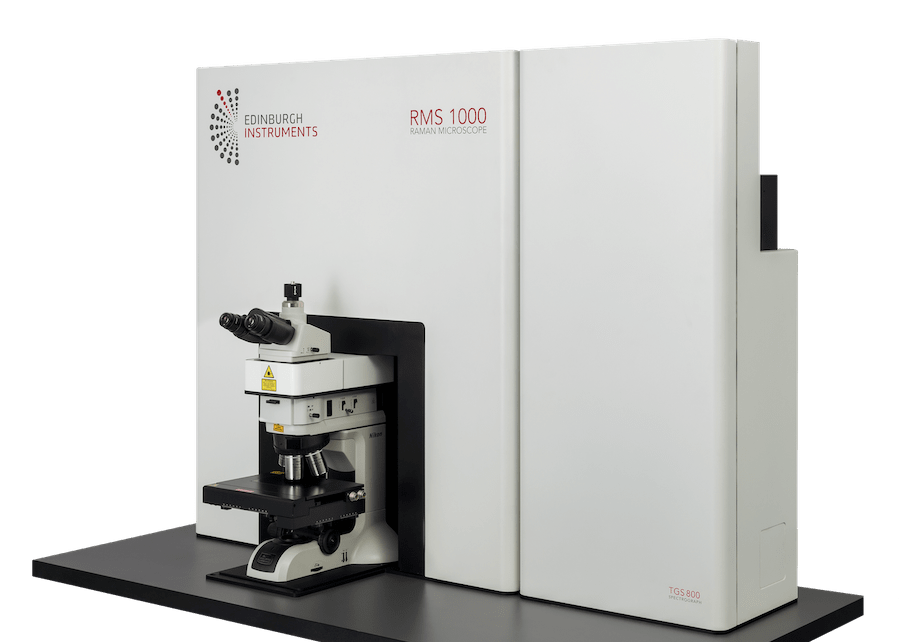 Edinburgh Instruments is delighted to announce the launch of the new RMS1000 Raman Microscope designed and manufactured at their global headquarters in Scotland. The RMS1000 Raman Microscope is an open architecture, research grade confocal Raman Microscope. It has been designed to be adapted to almost any modern, state-of-the-art Raman application. This high-end research tool has been built with no compromises; resulting in a system that stands alone in both specification and ease of use....