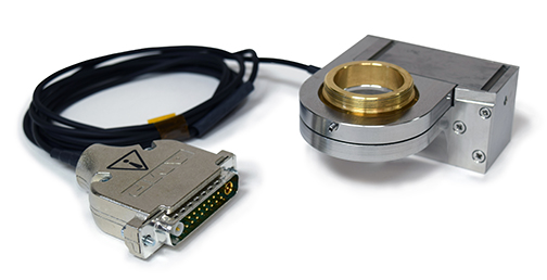
Prior Scientific is proud to announce the release of the NanoScan OP400, a piezo based objective scanner. Prior Scientific acquired Queensgate in 2018 and the NanoScan OP400 combines Prior’s expertise in delivering microscopy solutions with Queensgate’s market leading nanopositioning technology. The NanoScan OP400 provides the fastest step and settle time of any objective positioner available....
Analytik Ltd. has launched a range of benchtop Time-Domain Nuclear Magnetic Resonance (TD-NMR) systems. Applications that yield valuable data using TD-NMR include solid fat content and oil seed analysis in the food industry, obesity research and MRI contrast agents in the medical / pharmaceutical industry and a growing number of measurements in the chemical and polymer sectors...
Deben, a leading provider of in-situ testing stages together with innovative accessories and components for microscopy, reports on how Bristol Composites Institute of University of Bristol use the Deben 200N in-situ tensile stage to study Advanced Composite Materials for Aerospace applications. The Bristol Composites Institute (ACCIS) is a department of the University of Bristol looking at materials...
HORIBA UK Ltd, Medical announces the publication of scientific studies which demonstrate the excellent performance of its new HELO high throughput fully automated haematology platform on body fluid and pathological samples. HORIBA’s Yumizen® H2500 and H1500 automated haematology analysers within the HELO platform deliver enhanced precision for complete blood counts and white blood cell....
Merck, a leading science and technology company, presented Professor David Alsteens (33), Catholic University of Louvain (UCLovain), Louvain-la-Neuve, Belgium with the 2019 Heinrich Emanuel Merck Award for Analytical Science. The award ceremony took place during the analytical conference Euroanalysis at Istanbul University in Turkey. “With his groundbreaking investigations revealing the molecular mechanisms established by viruses to hijack the cellular barrier and enter the cell....
Bruker Corporation has announced the world’s first 1.2 GHz high-resolution, protein nuclear magnetic resonance (NMR) data. Two 1.2 GHz superconducting magnets have now reached full field at Bruker’s Swiss magnet factory, setting the world record for stable, homogeneous NMR magnets for high-resolution and solid-state protein NMR applications in structural biology...
Bruker has announced the European launch of the Fourier 80 system, a next-generation, high-performance 80 MHz Fourier Transform nuclear magnetic resonance (FT-NMR) benchtop spectrometer. The Fourier 80 has been designed for organic or medicinal chemistry research, routine analysis, teaching or synthesis verification in any chemistry laboratory....
Cryo transfer can enable or improve the ability to collect atom probe data in difficult applications, like: Rapid oxidation studies (e.g. uranium, lithium, aluminum), Catalyst/reaction chamber studies of surface contamination, Characterization of hydrogen embrittlement of steel, Transport between various analysis modalities (e.g. FIB, TEM) under vacuum conditions and Analysis of "soft" (i.e. biological) materials...
The new lyophilized exosomes have various biology applications, such as assay calibration, control for exosome quantification, protein marker analysis, extraction and analysis of exosome nucleic acid, standardized positive controls for immunocapture performance evaluation, flow cytometry, and electron microscopy....
MR Solutions has received the prestigious Queen’s Award for Enterprise in recognition of the company’s innovative PET imaging technology for use in pre-clinical research. MR Solutions already holds Queens Awards for innovation in 2016 and for export achievement in 2017.
Agilent Technologies Inc.,has introduced a multimode real-time cell analyzer (RTCA)—the first of its kind—combining the best of non-invasive biosensor measurement with live cell imaging. "The xCELLigence RTCA eSight will revolutionize cell analysis in life science research," said Todd Christian, Agilent vice president, and general manager of the company's Cell Analysis Division...
Recently, a number of studies have been published demonstrating that label-free, 3-D imaging using holotomography (HT) microscopy enables researchers to observe morphological and chemical alterations of host cells due to the parasite infection without any transfection or dye staining. This powerful new tool allows parasites to be easily and quickly detected and monitored within the host cells and permits the intricacies of parasite infection mechanisms and the host cell/parasite life cycle to be studied....
Vision Engineering, a manufacturer of high quality visual inspection and measurement technologies, launches its latest, innovative flagship product Deep Reality Viewer (DRV-Z1) microscope. Designed at its Woking, UK HQ, the DRV-Z1 enables the user to view high definition 3D images under magnification without using a flat screen, or requiring operators to wear goggles or specialist glasses. Uniquely, by linking multiple DRV systems via wired or wireless technologies....
ZEISS introduces new capabilities for ZEISS ion beam microscopes, which cover advancements in analytics, tomography, sample preparation and data integrity. This brings new possibilities in engineering materials, energy materials, soft materials and geosciences covering megatrends in additive manufacturing, battery and photovoltaic research, building materials and nanomaterials....
At this year’s European Congress of Pathology (ECP), Olympus will demonstrate how its 100 years of optical experience and the latest in ergonomic microscope design work together to make pathologists’ lives easier. Olympus will be displaying the culmination of 100 years of expertise in optics at booth R43 of the ECP in Nice from 7 to 11 September 2019. The 4K UHD UC90 camera, the new high-performance X Line objectives and the ergonomic BX46 microscope have each been carefully designed to enhance pathology workflows.
Laboratories are nearly always short of space which means bulky or very heavy equipment can’t be accommodated or has to be housed away from the laboratory. The introduction of liquid helium free, pre-clinical, high field MRI scanners has been a boon to the world’s leading laboratories. The commercially available liquid helium free technology which was developed by MR Solutions is now available in a range of MRI, and multi-modality MRI scanners (with PET and SPECT modules) from 3T up to 9.4T....
EIKOS-UV is a workhorse Atom Probe Microscope with increased ease of use and a low cost of ownership. Utilizing standard microscopy sample preparation methods, it delivers nanoscale structural information, enabling new a understanding of materials for research and faster development of products for industrial applications....
Olympus’ Image of the Year Award recognizes the best in life science imaging worldwide. Participants can win a CX43 microscope with a DP27 digital camera, X Line objectives, or an OM-D E-M5 Mark II camera. Those interested in participating can enter until 31 January 2020 by uploading images at the website. Winners will be selected by a jury panel and announced in March 2020....
London Resin has developed and supplied acrylic resins for microscopists since 1980, and this acquisition will ensure continued manufacture and supply of the complete product range. London Resin’s specially formulated, high quality microscopy resins combine low viscosity, low toxicity and ease of use, and are considered the industry standard for many applications....
TTP Labtech Ltd, a global leader in the design and development of automated instrumentation and consumables for life science applications, has announced the introduction of chameleon, an industry-enabling sample-preparation system for cryo-electron microscopy (cryo-EM). The new system has been designed to enable consistent application of samples to high-quality foil grids for cryo-EM analysis, automating the current standard approach, which is a technically-demanding, largely manual process....
Today’s educational landscape is changing rapidly thanks to technological advances. Introducing wireless capabilities to science classrooms enables lecturers to teach and students to learn in an engaging digital environment that promotes teamwork. Equipped with a WLAN-capable Olympus EP50 camera, every microscope in a classroom becomes a wireless imaging system...
ImaginAb Inc., a clinical stage immuno-oncology imaging company, announced today that it has entered into a non-exclusive license and collaboration agreement with Roche. Under the terms of the agreement, Roche will use ImaginAb's novel minibody CD8 T cell imaging agent in immuno-oncology clinical trials for multiple types of cancers....

 Prior Scientific is proud to announce the release of the NanoScan OP400, a piezo based objective scanner. Prior Scientific acquired Queensgate in 2018 and the NanoScan OP400 combines Prior’s expertise in delivering microscopy solutions with Queensgate’s market leading nanopositioning technology. The NanoScan OP400 provides the fastest step and settle time of any objective positioner available....
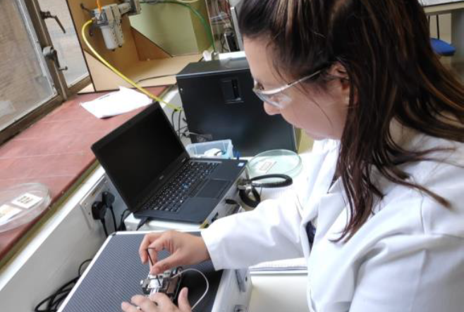 Deben, a leading provider of in-situ testing stages together with innovative accessories and components for microscopy, reports on how Bristol Composites Institute of University of Bristol use the Deben 200N in-situ tensile stage to study Advanced Composite Materials for Aerospace applications. The Bristol Composites Institute (ACCIS) is a department of the University of Bristol looking at materials...
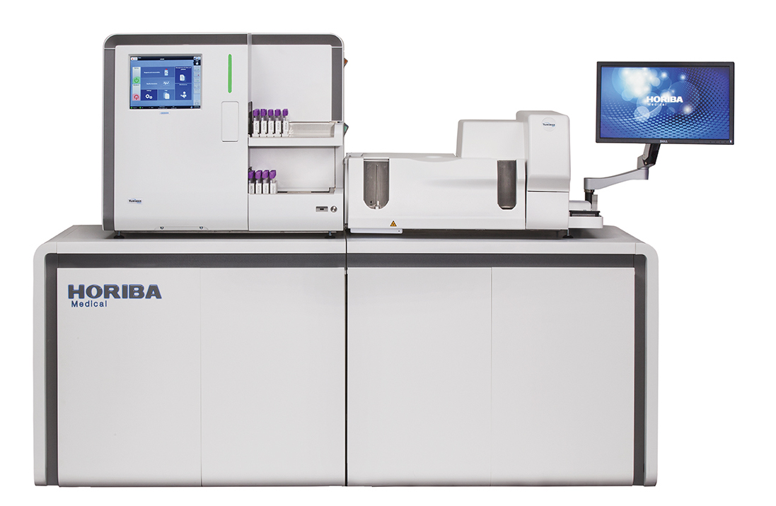 HORIBA UK Ltd, Medical announces the publication of scientific studies which demonstrate the excellent performance of its new HELO high throughput fully automated haematology platform on body fluid and pathological samples. HORIBA’s Yumizen® H2500 and H1500 automated haematology analysers within the HELO platform deliver enhanced precision for complete blood counts and white blood cell....
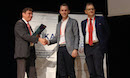 Merck, a leading science and technology company, presented Professor David Alsteens (33), Catholic University of Louvain (UCLovain), Louvain-la-Neuve, Belgium with the 2019 Heinrich Emanuel Merck Award for Analytical Science. The award ceremony took place during the analytical conference Euroanalysis at Istanbul University in Turkey. “With his groundbreaking investigations revealing the molecular mechanisms established by viruses to hijack the cellular barrier and enter the cell....
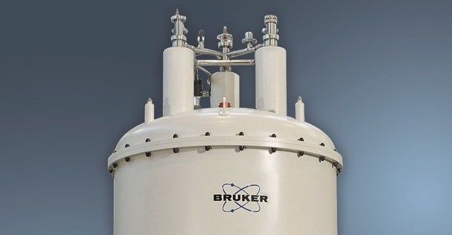 Bruker Corporation has announced the world’s first 1.2 GHz high-resolution, protein nuclear magnetic resonance (NMR) data. Two 1.2 GHz superconducting magnets have now reached full field at Bruker’s Swiss magnet factory, setting the world record for stable, homogeneous NMR magnets for high-resolution and solid-state protein NMR applications in structural biology...
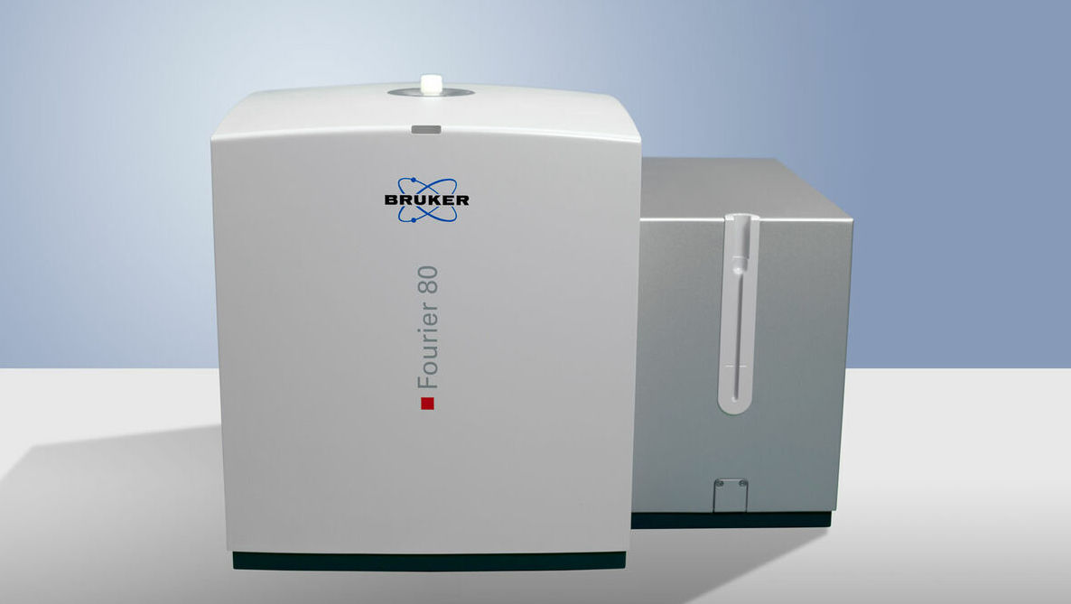 Bruker has announced the European launch of the Fourier 80 system, a next-generation, high-performance 80 MHz Fourier Transform nuclear magnetic resonance (FT-NMR) benchtop spectrometer. The Fourier 80 has been designed for organic or medicinal chemistry research, routine analysis, teaching or synthesis verification in any chemistry laboratory....
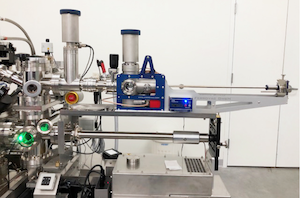 Cryo transfer can enable or improve the ability to collect atom probe data in difficult applications, like: Rapid oxidation studies (e.g. uranium, lithium, aluminum), Catalyst/reaction chamber studies of surface contamination, Characterization of hydrogen embrittlement of steel, Transport between various analysis modalities (e.g. FIB, TEM) under vacuum conditions and Analysis of "soft" (i.e. biological) materials...
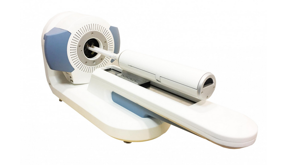 MR Solutions has received the prestigious Queen’s Award for Enterprise in recognition of the company’s innovative PET imaging technology for use in pre-clinical research. MR Solutions already holds Queens Awards for innovation in 2016 and for export achievement in 2017.
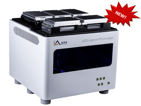 Agilent Technologies Inc.,has introduced a multimode real-time cell analyzer (RTCA)—the first of its kind—combining the best of non-invasive biosensor measurement with live cell imaging. "The xCELLigence RTCA eSight will revolutionize cell analysis in life science research," said Todd Christian, Agilent vice president, and general manager of the company's Cell Analysis Division...
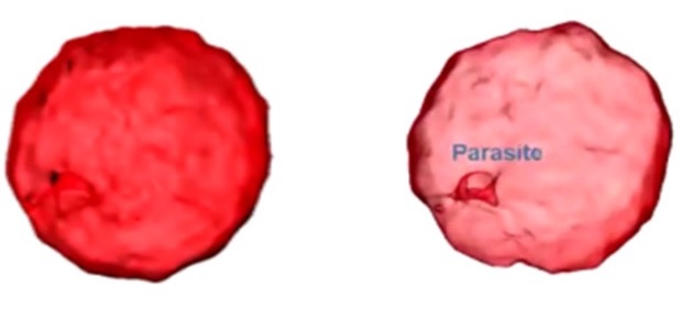 Recently, a number of studies have been published demonstrating that label-free, 3-D imaging using holotomography (HT) microscopy enables researchers to observe morphological and chemical alterations of host cells due to the parasite infection without any transfection or dye staining. This powerful new tool allows parasites to be easily and quickly detected and monitored within the host cells and permits the intricacies of parasite infection mechanisms and the host cell/parasite life cycle to be studied....
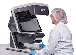 Vision Engineering, a manufacturer of high quality visual inspection and measurement technologies, launches its latest, innovative flagship product Deep Reality Viewer (DRV-Z1) microscope. Designed at its Woking, UK HQ, the DRV-Z1 enables the user to view high definition 3D images under magnification without using a flat screen, or requiring operators to wear goggles or specialist glasses. Uniquely, by linking multiple DRV systems via wired or wireless technologies....
 ZEISS introduces new capabilities for ZEISS ion beam microscopes, which cover advancements in analytics, tomography, sample preparation and data integrity. This brings new possibilities in engineering materials, energy materials, soft materials and geosciences covering megatrends in additive manufacturing, battery and photovoltaic research, building materials and nanomaterials....
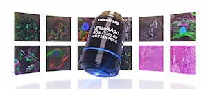 At this year’s European Congress of Pathology (ECP), Olympus will demonstrate how its 100 years of optical experience and the latest in ergonomic microscope design work together to make pathologists’ lives easier. Olympus will be displaying the culmination of 100 years of expertise in optics at booth R43 of the ECP in Nice from 7 to 11 September 2019. The 4K UHD UC90 camera, the new high-performance X Line objectives and the ergonomic BX46 microscope have each been carefully designed to enhance pathology workflows.
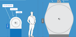 Laboratories are nearly always short of space which means bulky or very heavy equipment can’t be accommodated or has to be housed away from the laboratory. The introduction of liquid helium free, pre-clinical, high field MRI scanners has been a boon to the world’s leading laboratories. The commercially available liquid helium free technology which was developed by MR Solutions is now available in a range of MRI, and multi-modality MRI scanners (with PET and SPECT modules) from 3T up to 9.4T....
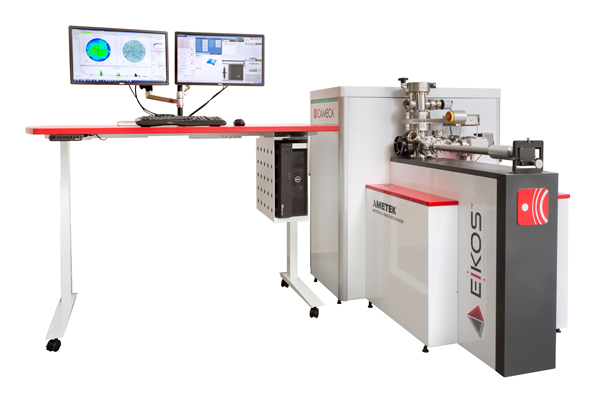 EIKOS-UV is a workhorse Atom Probe Microscope with increased ease of use and a low cost of ownership. Utilizing standard microscopy sample preparation methods, it delivers nanoscale structural information, enabling new a understanding of materials for research and faster development of products for industrial applications....
 Olympus’ Image of the Year Award recognizes the best in life science imaging worldwide. Participants can win a CX43 microscope with a DP27 digital camera, X Line objectives, or an OM-D E-M5 Mark II camera. Those interested in participating can enter until 31 January 2020 by uploading images at the website. Winners will be selected by a jury panel and announced in March 2020....
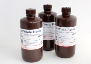 London Resin has developed and supplied acrylic resins for microscopists since 1980, and this acquisition will ensure continued manufacture and supply of the complete product range. London Resin’s specially formulated, high quality microscopy resins combine low viscosity, low toxicity and ease of use, and are considered the industry standard for many applications....
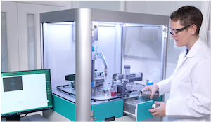 TTP Labtech Ltd, a global leader in the design and development of automated instrumentation and consumables for life science applications, has announced the introduction of chameleon, an industry-enabling sample-preparation system for cryo-electron microscopy (cryo-EM). The new system has been designed to enable consistent application of samples to high-quality foil grids for cryo-EM analysis, automating the current standard approach, which is a technically-demanding, largely manual process....
 Today’s educational landscape is changing rapidly thanks to technological advances. Introducing wireless capabilities to science classrooms enables lecturers to teach and students to learn in an engaging digital environment that promotes teamwork. Equipped with a WLAN-capable Olympus EP50 camera, every microscope in a classroom becomes a wireless imaging system...
 ImaginAb Inc., a clinical stage immuno-oncology imaging company, announced today that it has entered into a non-exclusive license and collaboration agreement with Roche. Under the terms of the agreement, Roche will use ImaginAb's novel minibody CD8 T cell imaging agent in immuno-oncology clinical trials for multiple types of cancers....