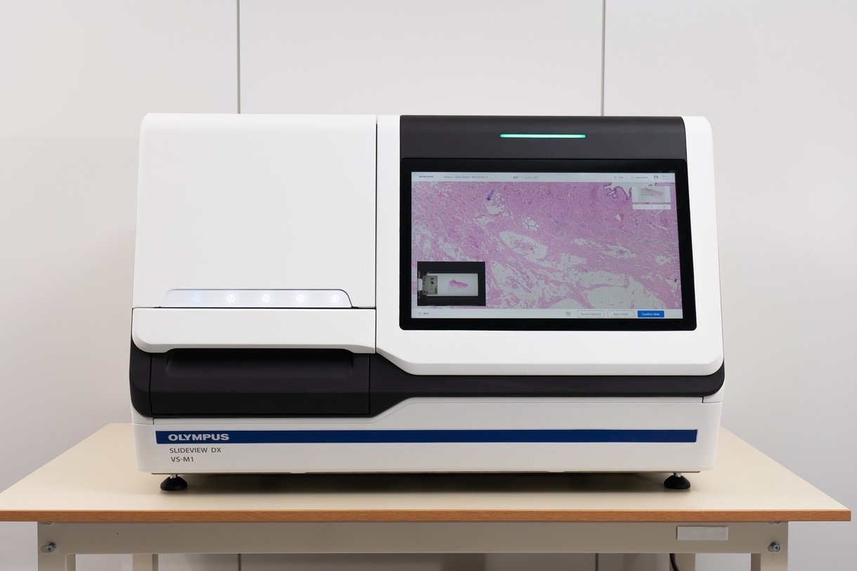
The new Evident SLIDEVIEW™ DX VS-M1 whole slide imaging system is a digital pathology solution that delivers high-quality slide images at high speed, helping pathologists make diagnoses quickly and efficiently. Built with renowned optics and advanced digital technology, the SLIDEVIEW DX slide scanner is a complete digital pathology solution. The SLIDEVIEW DX scanner provides microscope-quality images onscreen...
Digital Surf announce the release of the tenth major version of the company’s renowned Mountains® software analysis platform for surface metrology & microscopy, trusted by 50+ leading instrument manufacturers and 22,000+ users. Version 10 highlights include: A new product family for users of light microscopes, MountainsImage®, providing a complete toolkit for pre-processing and analyzing image data...
ZEISS Volutome is an end-to-end solution for serial block-face imaging from hardware to software including image processing, segmentation, and visualization. The ultramicrotome can be easily replaced by a conventional SEM stage, converting the 3D FE-SEM into a standard FE-SEM, making the system adaptable to a multi-purpose environment...
Vision Engineering, the world-leading provider of innovative inspection, metrology, and digital 3D visualisation solutions, today announced the launch of Mantis 3rd Gen, the latest addition to its best-selling and award-winning range of ergonomic optical stereo microscopes. Mantis is in use in tens of thousands of R&D, manufacturing and analytical sites around the world. Mantis 3rd Gen incorporates the latest developments in optics, digital cameras and fully adjustable LED lighting, to keep Mantis at the forefront of stereo imaging.
Revvity, Inc. announce that its EUROIMMUN business has launched the UNIQO 160 (CE-IVDR), an automated indirect immunofluorescence test (IIFT) system for autoimmune disease diagnostics. Now available in countries accepting the CE mark, this all-in-one solution increases the efficiency of the entire IIFT process, encompassing sample preparation, incubation, washing and mounting of slides as well as image acquisition and analysis...
ForceRobot 400 Enables High-Throughput Force Measurements for Single Molecule Research. Bruker has announced the release of the ForceRobot® 400 BioAFM, which marks a new milestone in force measurement capabilities. It sets new standards in automation, generating over 250,000 force curves per day to deliver the statistically significant datasets required for demanding discovery and preclinical research...
Have you read a ton of articles on phase contrast microscopy, but still can’t get the full picture? Well then, watch the second movie in the mini-movie series, “Microscopy With ibidi,” for a simple explanation of how this microscopy technique works. In this short movie, we will show you why this microscopy technique is so important for scientists. and how phase contrast makes almost transparent cells clearly visible...
An international team of scientists from The Institute of Cancer Research, London, and the Max Planck Institute for Multidisciplinary Sciences in Germany employed state-of-the-art biochemical and cryo-electron microscopy (cryo-EM) methods to study the spliceosome in intricate detail and answer long standing questions about how it works. The spliceosome operates like a nanobot, processing RNA – genetic instructions copied from DNA – in a key step to allow the building of complex proteins...
As the second leading cause of death worldwide, cancer has touched the lives of most Americans. In an effort to improve treatments for patients on Earth by better understanding how cancer spreads, a team of researchers is sending cancer organoids to space in an investigation sponsored by the International Space Station (ISS) National Laboratory. In collaboration with Axiom Space, researchers from the University of California San Diego and the Sanford Stem Cell Institute, will study the properties of cancer stem cells...
A new method of controlling the shape of tiny particles about one tenth of the width of human hair could make the technology that powers our daily lives more stable and more efficient, scientists claim. The process, which transforms the structure of microscopic semiconductor materials known as quantum dots, provides industry with opportunities to optimise optoelectronics, energy harvesting, photonics, and biomedical imaging technologies, according to the Cardiff University-led team...
Neu-tec Group Inc, in North America, is pleased to announce its new distribution partnership with the DK based Biosense Solutions. The oCelloscope technology is a unique live-cell imaging system for sensitive and detailed monitoring of biological growth and development. It uniquely tracks the growth of microorganisms in multi-well plates by combining microscopic imaging with automated time lapse photography...
Deluge of entries ensures one of the Society’s biggest and best ever competitions. This year’s RMS Scientific Imaging Competition has triggered a fantastic response from microscopists around the world – with 198 images submitted across seven different award categories. With the submissions deadline now expired, the competition judges have been left with the tricky task of whittling down a shortlist of images to be displayed at mmc2023, where the final decisions on the winners will be made...
TESCAN ORSAY HOLDING a.s. announces the first installation of its UniTOM XL with the new Spectral CT capability at University of Pau’s DMEX Centre for X-ray Imaging, Pau, France. Spectral CT provides chemical information at any point inside a sample, which is not possible using micro-CT alone. With Spectral CT scientists can now see the most subtle changes in material composition and purity, and low contrast materials, such as polymers, can be differentiated from each other...
England’s first centre of its kind is set to make significant improvements in cancer diagnosis and treatment – by combining pioneering digital imaging with artificial intelligence. The Royal Marsden NHS Foundation Trust and its academic research partner The Institute of Cancer Research, London, have today announced the opening of their new joint Integrated Pathology Unit (IPU)...
The ioLight compact inverted microscope is the latest innovation from ioLight. The inverted microscope wraps the microscope in a sealed package for use inside CO2 incubators in biological laboratories. The sealed case is resistant to damp acidic atmospheres and means that you can monitor experiments without opening the door or disrupting your cells. An external tablet can be used to view images remotely...
Indica Labs, an industry leader in quantitative digital pathology and image management innovations, and Lunit, a leading medical AI software platform company focused on cancer biomarkers announced an agreement to provide a fully interoperable solution between Indica Labs’ HALO AP® image management software platform and Lunit’s suite of AI pathology products. The combined solution is already in use at Guardant Health, a leading precision oncology company...
Get Better Images from Deep within Your Sample. The new SLIDEVIEW™ VS200 v. 4.1 software supports a host of new features, including the SILA optical sectioning device that enables you to obtain high-contrast images from deep within your sample. The SILA optical sectioning device uses speckle illumination combined with HiLo microscopy to achieve high-contrast images. This camera-based technology quickly captures two illuminated images that are then mathematically processed to remove out-of-focus light...
Prof. Dr. Immanuel Bloch, who is considered a leading quantum physicist, is to be commended with the ZEISS Research Award for his outstanding research in the field of quantum simulation using ultracold atoms. The company has been recognizing outstanding research in optics and photonics since 1990. The ceremony will take place at the Deutsches Museum in Munich on 26 June 2023. Three young scientists from Germany, Austria and Switzerland will also receive awards. They will receive the Carl Zeiss Award for Young Researchers...
Xyall and Indica Labs have entered into a global strategic partnership designed to bridge the gap between histology and molecular pathology. It unites Xyall’s unique automated tissue dissection solutions with Indica Labs’ AI-powered, diagnostic digital pathology platform. Selecting tumor tissue is at the heart of molecular diagnostic testing. The collaboration will enable molecular laboratories to benefit from a fully digital and automated workflow for tissue macro dissection for molecular diagnostic testing...
ibidi Glass Bottom µ-Plates With 384 Wells are now 30% off* until the end of June! With their highest optical quality coverslip, the µ-Plates enable high throughput screenings for many applications, such as Widefield fluorescence imaging and confocal microscopy, Total internal reflection fluorescence (TIRF) and Super-resolution microscopy (STED, SIM, (F)PALM, (d)STORM)...
CytoCHEK SPAchip® assay kits are novel fluorescence cell-based assays developed by A4cell that bring together the fields of nanotechnology and cell biology. SPAchips are composed of fluorescently labelled silicon microparticles which can be internalized inside single cells for lengthy periods, providing highly accurate data through long-term cell monitoring. CytoCHECK SPAchip® Detection Kits allow measurement of intracellular calcium or pH levels by changes in fluorescence intensity...
Transition metal dichalcogenides (TMDs) are a class of 2D materials which have unique optical and electronic properties. They are semiconductors with the chemical structure MX2 where M is a transition metal and X a chalcogen. After the isolation of graphene layers, research into alternative 2D layered materials which offer the same, or better, properties has surged...


 The new Evident SLIDEVIEW™ DX VS-M1 whole slide imaging system is a digital pathology solution that delivers high-quality slide images at high speed, helping pathologists make diagnoses quickly and efficiently. Built with renowned optics and advanced digital technology, the SLIDEVIEW DX slide scanner is a complete digital pathology solution. The SLIDEVIEW DX scanner provides microscope-quality images onscreen...
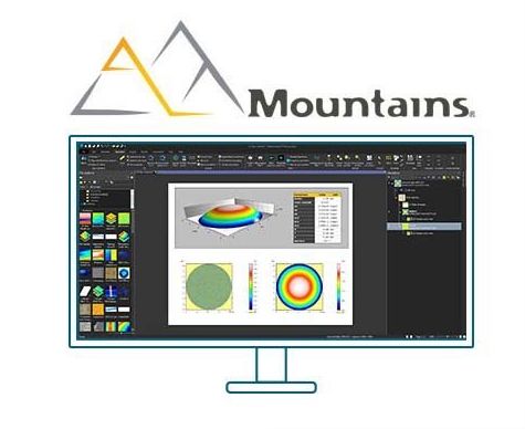 Digital Surf announce the release of the tenth major version of the company’s renowned Mountains® software analysis platform for surface metrology & microscopy, trusted by 50+ leading instrument manufacturers and 22,000+ users. Version 10 highlights include: A new product family for users of light microscopes, MountainsImage®, providing a complete toolkit for pre-processing and analyzing image data...
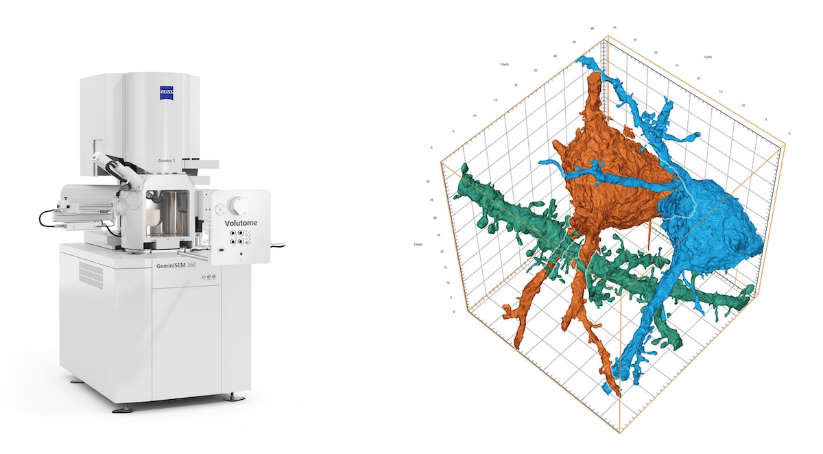 ZEISS Volutome is an end-to-end solution for serial block-face imaging from hardware to software including image processing, segmentation, and visualization. The ultramicrotome can be easily replaced by a conventional SEM stage, converting the 3D FE-SEM into a standard FE-SEM, making the system adaptable to a multi-purpose environment...
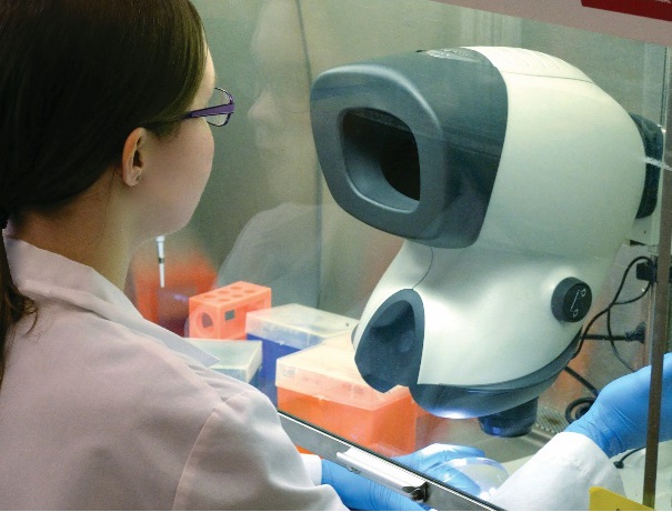 Vision Engineering, the world-leading provider of innovative inspection, metrology, and digital 3D visualisation solutions, today announced the launch of Mantis 3rd Gen, the latest addition to its best-selling and award-winning range of ergonomic optical stereo microscopes. Mantis is in use in tens of thousands of R&D, manufacturing and analytical sites around the world. Mantis 3rd Gen incorporates the latest developments in optics, digital cameras and fully adjustable LED lighting, to keep Mantis at the forefront of stereo imaging.
 Revvity, Inc. announce that its EUROIMMUN business has launched the UNIQO 160 (CE-IVDR), an automated indirect immunofluorescence test (IIFT) system for autoimmune disease diagnostics. Now available in countries accepting the CE mark, this all-in-one solution increases the efficiency of the entire IIFT process, encompassing sample preparation, incubation, washing and mounting of slides as well as image acquisition and analysis...
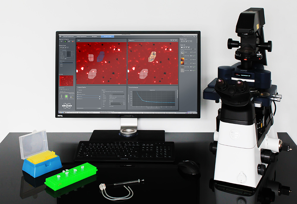 ForceRobot 400 Enables High-Throughput Force Measurements for Single Molecule Research. Bruker has announced the release of the ForceRobot® 400 BioAFM, which marks a new milestone in force measurement capabilities. It sets new standards in automation, generating over 250,000 force curves per day to deliver the statistically significant datasets required for demanding discovery and preclinical research...
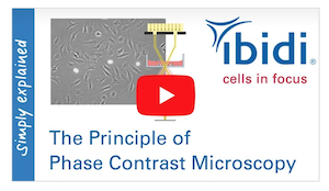 Have you read a ton of articles on phase contrast microscopy, but still can’t get the full picture? Well then, watch the second movie in the mini-movie series, “Microscopy With ibidi,” for a simple explanation of how this microscopy technique works. In this short movie, we will show you why this microscopy technique is so important for scientists. and how phase contrast makes almost transparent cells clearly visible...
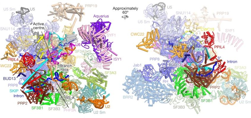 An international team of scientists from The Institute of Cancer Research, London, and the Max Planck Institute for Multidisciplinary Sciences in Germany employed state-of-the-art biochemical and cryo-electron microscopy (cryo-EM) methods to study the spliceosome in intricate detail and answer long standing questions about how it works. The spliceosome operates like a nanobot, processing RNA – genetic instructions copied from DNA – in a key step to allow the building of complex proteins...
 As the second leading cause of death worldwide, cancer has touched the lives of most Americans. In an effort to improve treatments for patients on Earth by better understanding how cancer spreads, a team of researchers is sending cancer organoids to space in an investigation sponsored by the International Space Station (ISS) National Laboratory. In collaboration with Axiom Space, researchers from the University of California San Diego and the Sanford Stem Cell Institute, will study the properties of cancer stem cells...
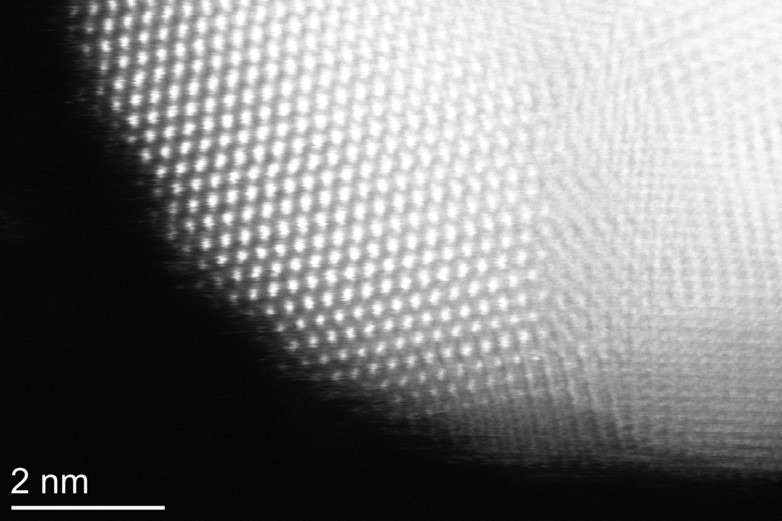 A new method of controlling the shape of tiny particles about one tenth of the width of human hair could make the technology that powers our daily lives more stable and more efficient, scientists claim. The process, which transforms the structure of microscopic semiconductor materials known as quantum dots, provides industry with opportunities to optimise optoelectronics, energy harvesting, photonics, and biomedical imaging technologies, according to the Cardiff University-led team...
 Neu-tec Group Inc, in North America, is pleased to announce its new distribution partnership with the DK based Biosense Solutions. The oCelloscope technology is a unique live-cell imaging system for sensitive and detailed monitoring of biological growth and development. It uniquely tracks the growth of microorganisms in multi-well plates by combining microscopic imaging with automated time lapse photography...
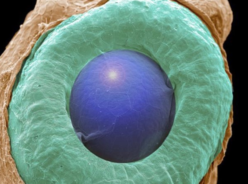 Deluge of entries ensures one of the Society’s biggest and best ever competitions. This year’s RMS Scientific Imaging Competition has triggered a fantastic response from microscopists around the world – with 198 images submitted across seven different award categories. With the submissions deadline now expired, the competition judges have been left with the tricky task of whittling down a shortlist of images to be displayed at mmc2023, where the final decisions on the winners will be made...
 TESCAN ORSAY HOLDING a.s. announces the first installation of its UniTOM XL with the new Spectral CT capability at University of Pau’s DMEX Centre for X-ray Imaging, Pau, France. Spectral CT provides chemical information at any point inside a sample, which is not possible using micro-CT alone. With Spectral CT scientists can now see the most subtle changes in material composition and purity, and low contrast materials, such as polymers, can be differentiated from each other...
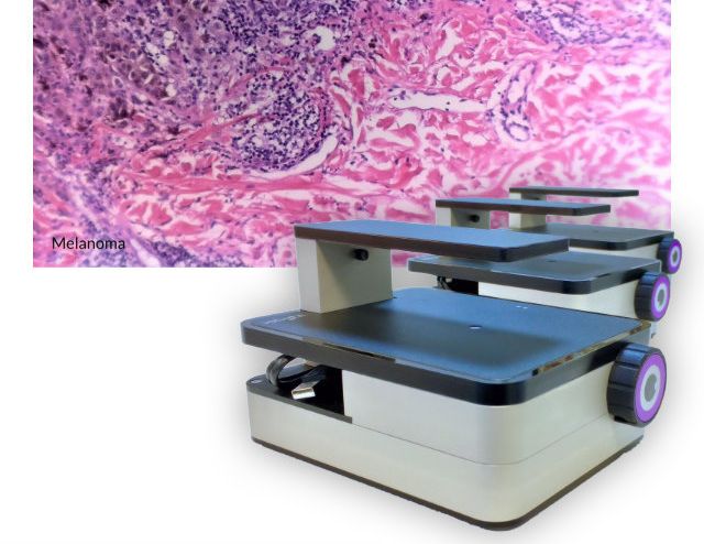 The ioLight compact inverted microscope is the latest innovation from ioLight. The inverted microscope wraps the microscope in a sealed package for use inside CO2 incubators in biological laboratories. The sealed case is resistant to damp acidic atmospheres and means that you can monitor experiments without opening the door or disrupting your cells. An external tablet can be used to view images remotely...
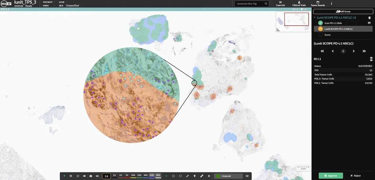 Indica Labs, an industry leader in quantitative digital pathology and image management innovations, and Lunit, a leading medical AI software platform company focused on cancer biomarkers announced an agreement to provide a fully interoperable solution between Indica Labs’ HALO AP® image management software platform and Lunit’s suite of AI pathology products. The combined solution is already in use at Guardant Health, a leading precision oncology company...
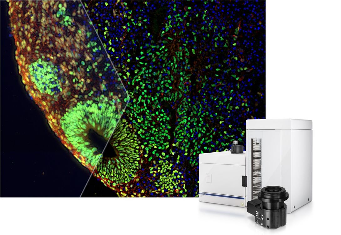 Get Better Images from Deep within Your Sample. The new SLIDEVIEW™ VS200 v. 4.1 software supports a host of new features, including the SILA optical sectioning device that enables you to obtain high-contrast images from deep within your sample. The SILA optical sectioning device uses speckle illumination combined with HiLo microscopy to achieve high-contrast images. This camera-based technology quickly captures two illuminated images that are then mathematically processed to remove out-of-focus light...
 Prof. Dr. Immanuel Bloch, who is considered a leading quantum physicist, is to be commended with the ZEISS Research Award for his outstanding research in the field of quantum simulation using ultracold atoms. The company has been recognizing outstanding research in optics and photonics since 1990. The ceremony will take place at the Deutsches Museum in Munich on 26 June 2023. Three young scientists from Germany, Austria and Switzerland will also receive awards. They will receive the Carl Zeiss Award for Young Researchers...
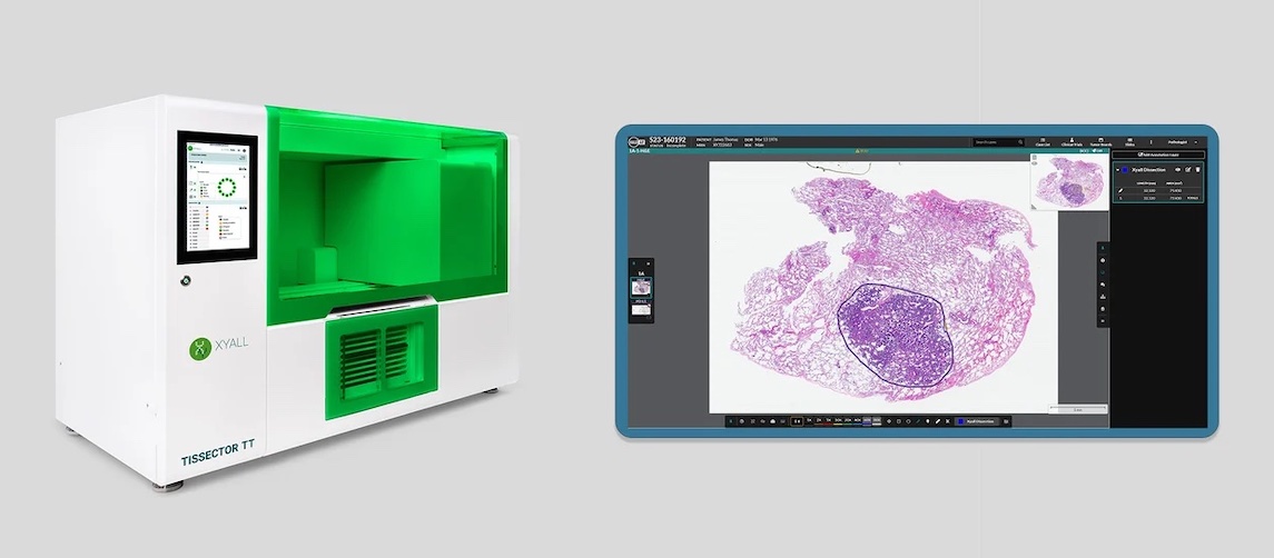 Xyall and Indica Labs have entered into a global strategic partnership designed to bridge the gap between histology and molecular pathology. It unites Xyall’s unique automated tissue dissection solutions with Indica Labs’ AI-powered, diagnostic digital pathology platform. Selecting tumor tissue is at the heart of molecular diagnostic testing. The collaboration will enable molecular laboratories to benefit from a fully digital and automated workflow for tissue macro dissection for molecular diagnostic testing...
 ibidi Glass Bottom µ-Plates With 384 Wells are now 30% off* until the end of June! With their highest optical quality coverslip, the µ-Plates enable high throughput screenings for many applications, such as Widefield fluorescence imaging and confocal microscopy, Total internal reflection fluorescence (TIRF) and Super-resolution microscopy (STED, SIM, (F)PALM, (d)STORM)...
 CytoCHEK SPAchip® assay kits are novel fluorescence cell-based assays developed by A4cell that bring together the fields of nanotechnology and cell biology. SPAchips are composed of fluorescently labelled silicon microparticles which can be internalized inside single cells for lengthy periods, providing highly accurate data through long-term cell monitoring. CytoCHECK SPAchip® Detection Kits allow measurement of intracellular calcium or pH levels by changes in fluorescence intensity...
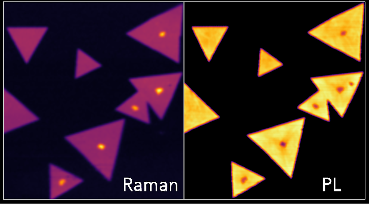 Transition metal dichalcogenides (TMDs) are a class of 2D materials which have unique optical and electronic properties. They are semiconductors with the chemical structure MX2 where M is a transition metal and X a chalcogen. After the isolation of graphene layers, research into alternative 2D layered materials which offer the same, or better, properties has surged...