Researchers have made a significant breakthrough in understanding how a family of viruses, including the norovirus, initiate infections. The new study, published today in Nature and led by the University of Glasgow, reveals the inner workings of the calicivirus family, which includes norovirus and sapoviruses – highly infectious viruses that can cause outbreaks of diarrhoea and vomiting. It is hoped this research may provide a new target for the development of antiviral drugs to prevent diseases like norovirus...
The powerful new software module ZEISS Labscope Teacher expands the existing ZEISS Labscope installation. It puts the lecturer in charge of all connected microscopes in the network of the digital classroom while they move freely around the classroom. It lets the teacher define working groups, send group-specific tasks, and share digital information such as documents or presentations, thereby fostering teamwork...
Prior Scientific Instruments Ltd marks its 100th anniversary of the company’s founding in 1919, a rare achievement in today’s business climate. “This is a proud time for the company and illustrates our resiliency and adaptability. We look forward to the next 100 years and the developments that we can bring to our markets”, said Thomas Freda, CEO of Prior Scientific. “Prior is a very different company from what it was 100, 50, or even 10 years ago....
Renishaw inVia™ microscope user, Prof. Carlos A. Carrero, heads a group that is developing advanced catalysts for the conversion of hydrocarbons into more valuable products at the Department of Chemical Engineering, Auburn University, USA. The Carrero group undertakes fundamental studies to improve and develop more efficient and sustainable catalytic processes. They aim to develop the best catalysts using in situ / operando Raman spectroscopy to gather molecular, structural, and mechanistic insights....
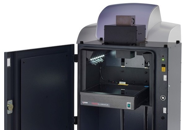
With years of experience in the pharmaceutical and life science sector, Creative Diagnostics recently launches a wide range of fluorescent silica particles with a high amount of covalently bound fluorescence dye in the silica matrix. These new fluorescent silica nanoparticles are available with red fluorescence, green fluorescence and blue fluorescence, which can be applied in fluorescent imaging and multimodal imaging, drug and gene delivery, and theranostics platform....
ZEISS is introducing ZEISS Elyra 7 with Lattice SIM, a new flexible platform for fast and gentle 3D superresolution. Lattice SIM expands the possibilities of structured illumination microscopy (SIM): illuminating the sample with a lattice pattern rather than grid lines gives higher contrast and allows a more robust image reconstruction. Scientists can use 2x higher sampling efficiency to lower the illumination dosage to observe fast cellular processes in superresolution....
The new Renishaw RA816 Biological Analyser is a compact benchtop Raman imaging system, designed for biological and clinical research. This easy-to-use instrument enables the rapid collection of detailed information from a range of biological samples, including tissue and biofluids. Detailed biochemical information is revealed from biological samples. The Renishaw RA816 Biological Analyser enables biologists and clinicians to...
ZEISS announces its biggest new release of the ZEISS Mineralogic software at the Process Mineralogy '18 conference in Cape Town, South Africa. This is the 7th instalment of ZEISS Mineralogic since the software was brought to the market in July 2014 and represents a significant advancement in both features and productivity. The software is already well known for providing quantitative mineralogy with the...
HT-2 microscope facilitates three-dimensional fluorescence and optical diffraction tomography of live cells with minimal stress on cells. The HT-2 from Tomocube is the world’s first microscope to combine both holotomography and 3D fluorescence imaging into one unit. Capable of simultaneously capturing high resolution 3D optical diffraction tomography and 3D fluorescence images, the new microscope enables long-term tracking of specific targets in live cells while...
Featuring a Super-sensitive Lens for Quick, Accurate Imaging of all Gels and Blots. Syngene, a world-leading manufacturer of image analysis solutions, is delighted to announce its new generation G:BOX Chemi XX6/XX9 multi-application imaging systems are now available. Featuring the latest, ultra-sensitive, fast capture lens, these systems ensure scientists can have total confidence that their gel and blot results are accurate and publication-ready...
The breath-taking winners of the 44th annual competition unveil the microscopic beauty hidden from the naked eye. Nikon Instruments Inc. has announced the winners of the forty-fourth annual Nikon Small World Photomicrography Competition. First place was awarded to Emirati photographer Yousef Al Habshi, who sees the eyes as the windows to stunning insect artwork and research. The 2018 winning image captures part of the compound eyes and surrounding greenish scales of an Asian Red Palm weevil....
The Olympus high-sensitivity SpinSR10 super resolution microscope combines fast 0.005 seconds/frame acquisition speed and 120-nanometer super resolution with improvements that provide clearer images and three times the brightness of the standard SpinSR10 model. Super resolution microscopes are used in medical and biological research to observe the internal structure of cells in fine detail, and brightness and clarity are essential to image quality....
New microscopy technologies, such as light sheet fluorescence microscopy (LSFM) or clearing methods, allow the imaging of large samples at high resolution or high frame rates. Handling, processing, and analyzing these multi-terabyte data sets has become increasingly difficult for scientists, e.g. in developmental biology and neuroscience. New tools are needed to overcome these challenges. Therefore, EISS has teamed up with arivis AG to offer complete solutions from initial image acquisition to final results...
MR Solutions continues to expand its range of preclinical scanners with the first showing of its elegant bench top CT scanner. The scanner which was displayed for the first time at the EANM congress in Dusseldorf in October can be fitted with either a PET or SPECT clip-on. MR Solutions now has four CT models from rodent imaging up to the newly announced Large Field of View with a 24cm field. The clip-on PET and SPECT scanners, which can also be used as standalone scanners, and...
At the Annual Meeting of the Society for Neuroscience 2018 (Nov 03 – 07), ZEISS presented new workflows in collaboration with Inscopix (Palo Alto, CA) where ZEISS’s Airyscan confocal imaging technology is leveraged with the Inscopix freely behaving microscopy systems. The joint workflow development utilizes the increased spatial resolution, signal-to-noise and sectioning of ZEISS Airyscan to further inform the freely behaving signaling data collected with the Inscopix system....
AMSBIO has expanded its range of high quality FFPE cancer cell line controls for immunohistochemistry (IHC), in situ hybridization (ISH) and Next Generation Sequencing (NGS) applications. Variations in FFPE tissue sample quality and experimental setup can lead to misleading results without reliable controls. Traditional cell or tissue-based control specimens often present intra specimen...
With the concerted efforts of its scientists, BOC Sciences recently released fluorescent labeling service to facilitate the research work in the scientific community, adding to its already existing service portfolio encompassing custom synthesis, process R&D, bioconjugation, carbohydrate synthesis, and more. Fluorescent labelling is the process of covalently binding fluorescent dyes to biomolecules such as nucleic acids or proteins so that they can be visualized by fluorescence imaging....
Global support for Holotomography Microscopy as company prepares for introduction of second microscope model. A comprehensive network of specialist imaging companies has been unveiled to support the sales and maintenance of Tomocube’s holotomography microscopes around the world. Covering North America, Europe and Asia, the 23-strong group is being launched as Tomocube prepares to introduce its second microscope model in less than three months....
Joint further development of 3D visualizations of multidimensional image data. ZEISS and arivis have now expanded and deepened their cooperation, which began in 2014. The companies are pursuing the common goal of enabling new findings in life sciences and in materials research by improving the presentation and analysis of digital data. Modern microscope systems with high-resolution optics record ever increasing amounts of data, which can only be displayed and analyzed with the help of advanced software...
BioTek has released a Peltier Cooling Modulefor the Cytation™ Cell Imaging Multi-Mode Readers. The compact module keeps internal temperature rise to less than one degree over ambient, regardless of fluctuations from external and internal factors. The module helps maintain and optimize stability for more consistent data in assays run at ambient temperature. The module also quickly reduces internal temperature after incubated assays for efficient transitions between multiple applications with different temperature requirements....
Leeds Teaching Hospitals NHS Trust and the University of Leeds have today announced a critical milestone in going digital, by scanning every glass slide they produce. The milestone represents a major step towards achieving faster and accurate diagnosis for cancer patients in the future. The Pathology Department, located in St. James’ Hospital in Leeds, is one of the largest in the UK processing over 1,000 pathology slides a day, and is now digitally scanning every slide thanks to their partnership with Leica Biosystems....
Discover the LabRAM Odyssey™ Anniversary Special Edition, fully loaded with our most popular innovations: Duoscan, ULF, SWIFT XS EMCCD, at an amazing price! Special 50 years anniversary series: true confocal Raman microscope enabling the most detailed images and analyses to be obtained with speed and confidence. Ideally suited for both micro and macro measurements, it offers advanced confocal imaging capabilities in 2D and 3D. With guaranteed high performance and intuitive simplicity, the LabRAM Odyssey™ is the ultimate instrument for Raman spectroscopy....


 Prior Scientific Instruments Ltd marks its 100th anniversary of the company’s founding in 1919, a rare achievement in today’s business climate. “This is a proud time for the company and illustrates our resiliency and adaptability. We look forward to the next 100 years and the developments that we can bring to our markets”, said Thomas Freda, CEO of Prior Scientific. “Prior is a very different company from what it was 100, 50, or even 10 years ago....
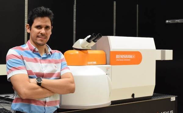 Renishaw inVia™ microscope user, Prof. Carlos A. Carrero, heads a group that is developing advanced catalysts for the conversion of hydrocarbons into more valuable products at the Department of Chemical Engineering, Auburn University, USA. The Carrero group undertakes fundamental studies to improve and develop more efficient and sustainable catalytic processes. They aim to develop the best catalysts using in situ / operando Raman spectroscopy to gather molecular, structural, and mechanistic insights....
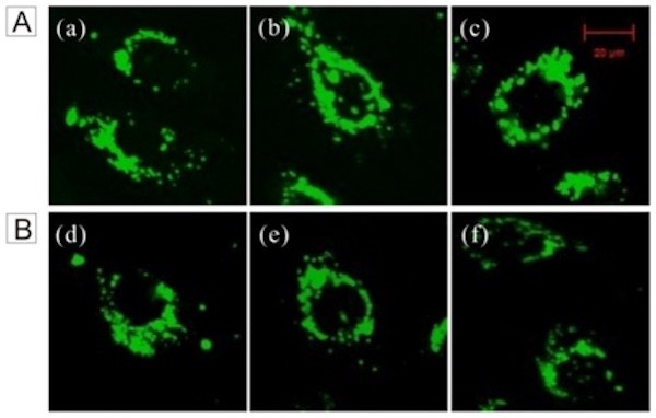
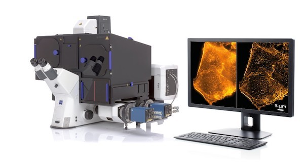 ZEISS is introducing ZEISS Elyra 7 with Lattice SIM, a new flexible platform for fast and gentle 3D superresolution. Lattice SIM expands the possibilities of structured illumination microscopy (SIM): illuminating the sample with a lattice pattern rather than grid lines gives higher contrast and allows a more robust image reconstruction. Scientists can use 2x higher sampling efficiency to lower the illumination dosage to observe fast cellular processes in superresolution....
 The new Renishaw RA816 Biological Analyser is a compact benchtop Raman imaging system, designed for biological and clinical research. This easy-to-use instrument enables the rapid collection of detailed information from a range of biological samples, including tissue and biofluids. Detailed biochemical information is revealed from biological samples. The Renishaw RA816 Biological Analyser enables biologists and clinicians to...
 ZEISS announces its biggest new release of the ZEISS Mineralogic software at the Process Mineralogy '18 conference in Cape Town, South Africa. This is the 7th instalment of ZEISS Mineralogic since the software was brought to the market in July 2014 and represents a significant advancement in both features and productivity. The software is already well known for providing quantitative mineralogy with the...
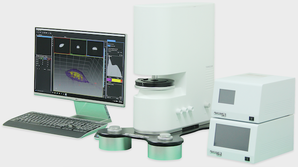
 Featuring a Super-sensitive Lens for Quick, Accurate Imaging of all Gels and Blots. Syngene, a world-leading manufacturer of image analysis solutions, is delighted to announce its new generation G:BOX Chemi XX6/XX9 multi-application imaging systems are now available. Featuring the latest, ultra-sensitive, fast capture lens, these systems ensure scientists can have total confidence that their gel and blot results are accurate and publication-ready...
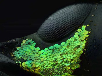 The breath-taking winners of the 44th annual competition unveil the microscopic beauty hidden from the naked eye. Nikon Instruments Inc. has announced the winners of the forty-fourth annual Nikon Small World Photomicrography Competition. First place was awarded to Emirati photographer Yousef Al Habshi, who sees the eyes as the windows to stunning insect artwork and research. The 2018 winning image captures part of the compound eyes and surrounding greenish scales of an Asian Red Palm weevil....
.jpg) The Olympus high-sensitivity SpinSR10 super resolution microscope combines fast 0.005 seconds/frame acquisition speed and 120-nanometer super resolution with improvements that provide clearer images and three times the brightness of the standard SpinSR10 model. Super resolution microscopes are used in medical and biological research to observe the internal structure of cells in fine detail, and brightness and clarity are essential to image quality....
The Olympus high-sensitivity SpinSR10 super resolution microscope combines fast 0.005 seconds/frame acquisition speed and 120-nanometer super resolution with improvements that provide clearer images and three times the brightness of the standard SpinSR10 model. Super resolution microscopes are used in medical and biological research to observe the internal structure of cells in fine detail, and brightness and clarity are essential to image quality....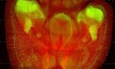 New microscopy technologies, such as light sheet fluorescence microscopy (LSFM) or clearing methods, allow the imaging of large samples at high resolution or high frame rates. Handling, processing, and analyzing these multi-terabyte data sets has become increasingly difficult for scientists, e.g. in developmental biology and neuroscience. New tools are needed to overcome these challenges. Therefore, EISS has teamed up with arivis AG to offer complete solutions from initial image acquisition to final results...
 MR Solutions continues to expand its range of preclinical scanners with the first showing of its elegant bench top CT scanner. The scanner which was displayed for the first time at the EANM congress in Dusseldorf in October can be fitted with either a PET or SPECT clip-on. MR Solutions now has four CT models from rodent imaging up to the newly announced Large Field of View with a 24cm field. The clip-on PET and SPECT scanners, which can also be used as standalone scanners, and...
 At the Annual Meeting of the Society for Neuroscience 2018 (Nov 03 – 07), ZEISS presented new workflows in collaboration with Inscopix (Palo Alto, CA) where ZEISS’s Airyscan confocal imaging technology is leveraged with the Inscopix freely behaving microscopy systems. The joint workflow development utilizes the increased spatial resolution, signal-to-noise and sectioning of ZEISS Airyscan to further inform the freely behaving signaling data collected with the Inscopix system....
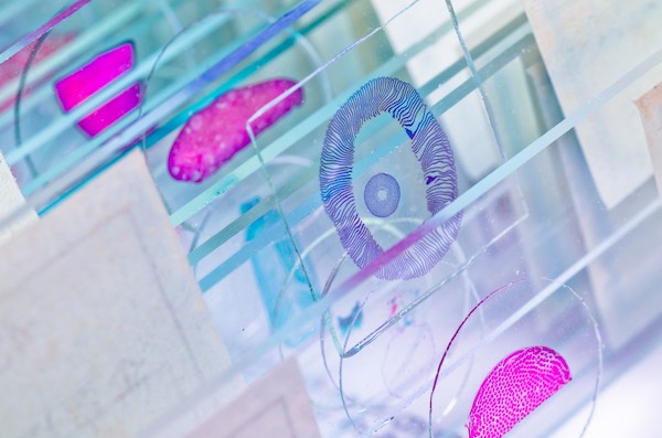 AMSBIO has expanded its range of high quality FFPE cancer cell line controls for immunohistochemistry (IHC), in situ hybridization (ISH) and Next Generation Sequencing (NGS) applications. Variations in FFPE tissue sample quality and experimental setup can lead to misleading results without reliable controls. Traditional cell or tissue-based control specimens often present intra specimen...
 With the concerted efforts of its scientists, BOC Sciences recently released fluorescent labeling service to facilitate the research work in the scientific community, adding to its already existing service portfolio encompassing custom synthesis, process R&D, bioconjugation, carbohydrate synthesis, and more. Fluorescent labelling is the process of covalently binding fluorescent dyes to biomolecules such as nucleic acids or proteins so that they can be visualized by fluorescence imaging....
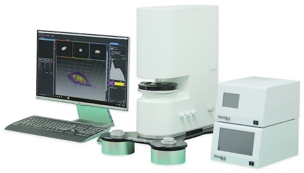 Global support for Holotomography Microscopy as company prepares for introduction of second microscope model. A comprehensive network of specialist imaging companies has been unveiled to support the sales and maintenance of Tomocube’s holotomography microscopes around the world. Covering North America, Europe and Asia, the 23-strong group is being launched as Tomocube prepares to introduce its second microscope model in less than three months....
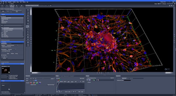 Joint further development of 3D visualizations of multidimensional image data. ZEISS and arivis have now expanded and deepened their cooperation, which began in 2014. The companies are pursuing the common goal of enabling new findings in life sciences and in materials research by improving the presentation and analysis of digital data. Modern microscope systems with high-resolution optics record ever increasing amounts of data, which can only be displayed and analyzed with the help of advanced software...
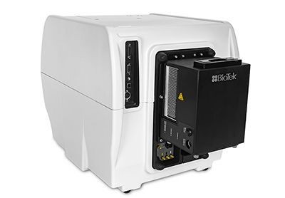 BioTek has released a Peltier Cooling Modulefor the Cytation™ Cell Imaging Multi-Mode Readers. The compact module keeps internal temperature rise to less than one degree over ambient, regardless of fluctuations from external and internal factors. The module helps maintain and optimize stability for more consistent data in assays run at ambient temperature. The module also quickly reduces internal temperature after incubated assays for efficient transitions between multiple applications with different temperature requirements....
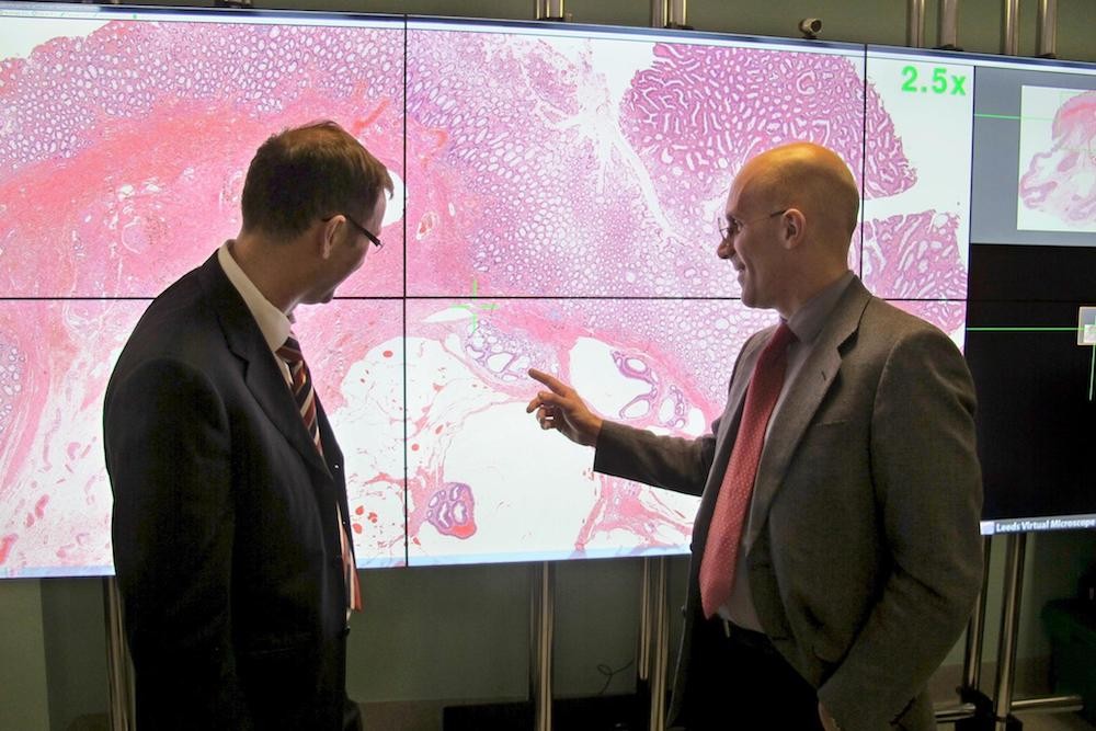 Leeds Teaching Hospitals NHS Trust and the University of Leeds have today announced a critical milestone in going digital, by scanning every glass slide they produce. The milestone represents a major step towards achieving faster and accurate diagnosis for cancer patients in the future. The Pathology Department, located in St. James’ Hospital in Leeds, is one of the largest in the UK processing over 1,000 pathology slides a day, and is now digitally scanning every slide thanks to their partnership with Leica Biosystems....
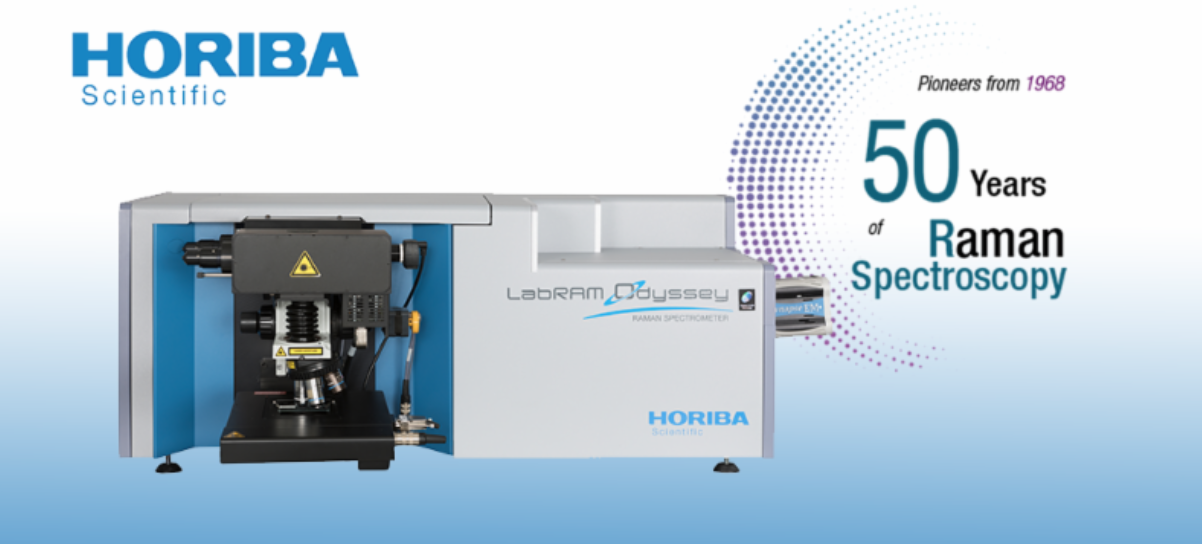 Discover the LabRAM Odyssey™ Anniversary Special Edition, fully loaded with our most popular innovations: Duoscan, ULF, SWIFT XS EMCCD, at an amazing price! Special 50 years anniversary series: true confocal Raman microscope enabling the most detailed images and analyses to be obtained with speed and confidence. Ideally suited for both micro and macro measurements, it offers advanced confocal imaging capabilities in 2D and 3D. With guaranteed high performance and intuitive simplicity, the LabRAM Odyssey™ is the ultimate instrument for Raman spectroscopy....