BioTek introduces the new AutoScratch™Wound Making Tool to facilitate two dimensional (2D) collective cell migration and invasion applications. With a simple pushbutton operation, the AutoScratch automatically creates consistent scratches of equivalent size and area in confluent cell monolayers grown in 24- or 96-well microplate format. This eliminates variability found in manual scratch methods and increases the reproducibility necessary to analyze and normalize results across subsequent assays....
In the electronics quality control (QC) process by which entities review the quality of all factors involved in production, a visual product inspection is often performed. Here, every product is examined visually, often using a microscope due to the miniaturization before the product is sold into the external market. Inspectors will be provided with lists and descriptions of unacceptable product defects such as cracks or surface blemishes. Using a digital microscope enables the inspector to faster detect product defects by checking the magnified boards in FULL HD image quality.
Ground-breaking technology unlocks rapid and simple label-free 3D and 4D live cell imaging. A novel optical microscope utilizing diffraction tomography to generate 3D holographic images of unlabelled live cells is now available from Tomocube. Called HT-1, the new microscope delivers quantitative nanoscale, real-time, dynamic images of individual living cells quickly and simply without any sample preparation...
Money under the microscope - Financial comparison website money.co.uk, teamed up with the London Metropolitan University to put money under the microscope to discover what’s really lurking in your wallet - the findings are not for the faint hearted. Dr Paul Matewele, Professor of Microbiology at London Met, and his students took 36 samples from a random selection of all denominations of coins and notes. The microbiologists studied the bacteria in a controlled lab environment over a period of 8 weeks....
FLIR Systems announces update version 4.40 of its popular thermal measurement, recording and analysis software - ResearchIR. ResearchIR 4.40 is a powerful, yet easy to-use thermal analysis software package for research grade uncooled and cooled cameras from FLIR Systems. The latest revision of this software suite provides highly intuitive advanced camera...
Phasefocus™ is delighted to announce the launch of a new corporate website. With a fresh look and feel, the new site has been designed to improve the end-user experience, providing simple, logical, navigation, allowing visitors to rapidly locate and access a wealth of product and application information from a single location. Visitors to the site can now browse the wide range of Livecyte™ applications, from where they can...
High resolution 3D block face imaging for biological samples with fast acquisition rates and minimal sample damage. In collaboration with the National Center for Microscopy and Imaging Research (NCMIR) at the University of California San Diego, ZEISS releases a new Focal Charge Compensation module for block face imaging with ZEISS GeminiSEM and 3View® from Gatan, Inc. Focal Charge Compensation...
Keynote speakers have been announced for HSI 2018, the 7th international conference on Hyperspectral Imaging & Applications, co-located with Photonex Europe taking place on 10th & 11th October at the Ricoh Arena, Coventry, UK. HSI 2018 brings together academic and industrial users to share their knowledge of innovations in hyperspectral imaging cameras, technology and applications....
Understanding the process of interaction between healthy and diseased cells provides a better understanding of the life processes of organisms, resulting in more effective medicines, new vaccines. In every successful relationship, communication is key. This can be among friends in a social setting, at home with family or in the workplace with colleagues....
Founder and Chairman, Dr David Taylor, of Guildford based MR Solutions has been shortlisted as an Innovation Entrepreneur of the Year for London and the South East in the 2018 NatWest Great British Entrepreneur Awards. The awards have had a record number of entries with businesses from across the south east vying for a place on the shortlist...
The University of Glasgow has opened an innovative new structural biology centre, home to a cutting-edge electron microscope – the first of its kind in Scotland – which will be used to image biological molecules at the atomic level. The technology will be used to support vital research into diseases posing the greatest threat to human and animal health, providing greater capabilities in areas such as vaccine development, cancer research, and drug design and discovery....
Tomocube’s revolutionary holotomography technique delivers 3D structural and chemical information quickly and simply without any labelling or pre-preparation. A spin-out company from the renowned Korea Advanced Institute of Science and Technology in Daejeon has been established to commercialize the revolutionary Holotomography (HT) microscopy developed...
Creative Diagnostics recently launches fluorescent gold nanorods, labeled with various fluorophores for multimodal imaging for the global life science marketplace, which can be applied in various research fields including drug delivery, photothermal therapy, multimodal imaging, dark-field scattering imaging, computed tomography, and so on.
Market leaders in temperature-controlled microscopy and leaders in sample characterization solutions, Linkam Scientific Instruments, reports on the research of Professor Xiaoming “Shawn” He of the Fischell Department of Bioengineering at the University of Maryland. Professor He's research is focused on developing multiscale biomaterials to engineer living cells and tissues for two applications: Cancer treatment and cell-based medicine...
A team of scientists at the Sainsbury Laboratory Cambridge University (SLCU) have discovered a rare mineral in alpine plants. Vaterite, a form (polymorph) of calcium carbonate, holds enticing potential as a new material for industrial and medical applications. This is the first time that vaterite has been found in plants. The discovery of vaterite was made through a collaboration between SLCU and Cambridge University Botanic Garden....
ZEISS presents the initial release of its cloud-based digital microscopy platform, known under the name APEER at the Microscopy & Microanalysis conference (M&M) in Baltimore, USA. Microscopy users will be able to automatically process images in the cloud by leveraging application workflows for 3D reconstructions, staining or segmenting...
ZEISS expands X-ray imaging portfolio with ZEISS Xradia Context microCT. Built on renowned ZEISS Xradia platform and field-convertible to ZEISS Xradia Versa X-ray microscope. ZEISS introduces a new X-ray imaging instrument, ZEISS Xradia Context microCT, a large field-of-view, non-destructive 3D X-ray microcomputed tomography system. ZEISS Xradia Context is built on time-tested ZEISS Xradia technology, reaping crossover benefits of years of...
CAMECA, a world leader in scientific instrumentation and metrology solutions, is pleased to announce the launch of SXFive-TACTIS, the newest addition to the CAMECA line of high-end microanalytical instruments, and the only Electron Probe Microanalyzer (EPMA) in the world with a touch-screen interface. “We are very proud to introduce the SXFive-TACTIS EPMA, which represents the latest evolution in our SXFive product line,” comments Sanjay Kamtekar...
First ZEISS ZEN Intellesis solution enables segmentation of correlative microscopy datasets. ZEISS recently announced ZEISS ZEN Intellesis, a new machine learning capability that enables researchers to perform advanced analysis of their imaging samples across multiple microscopy methods. The first algorithmic solution introduced by the...
Linkam Scientific Instruments, reports on the research of Dr Jamie Wilkinson of the Natural History Museum in London where he uses temperature-controlled microscopy to characterize mineral deposits. Dr Jamie Wilkinson is a Research Leader in Mineral Deposits in the Department of Earth Sciences at the Natural History Museum, London. He is a member of the London Centre for Ore Deposits and Exploration (LODE) Research Group...
JPK Instruments, a world-leading manufacturer of nanoanalytic instrumentation for research in life sciences and soft matter, reports on how the Nano Imaging Lab of the Swiss Nanoscience Institute uses JPK’s versatile NanoWizard® 4 AFM system. Dr Monica Schönenberger has developed a flexible, professional service unit for atomic force microscopy. As a member of the Swiss Nanoscience Institute (SNI)...
MR Solutions has installed Hawaii’s first preclinical cryogen-free MRI imaging system to help researchers improve the rate of kidney disease detection through research. This is very good news for Hawaii which has a very high incidence of kidney disease - one in seven residents on the island has chronic kidney disease....


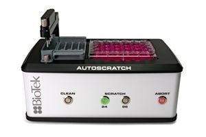 BioTek introduces the new AutoScratch™Wound Making Tool to facilitate two dimensional (2D) collective cell migration and invasion applications. With a simple pushbutton operation, the AutoScratch automatically creates consistent scratches of equivalent size and area in confluent cell monolayers grown in 24- or 96-well microplate format. This eliminates variability found in manual scratch methods and increases the reproducibility necessary to analyze and normalize results across subsequent assays....
 In the electronics quality control (QC) process by which entities review the quality of all factors involved in production, a visual product inspection is often performed. Here, every product is examined visually, often using a microscope due to the miniaturization before the product is sold into the external market. Inspectors will be provided with lists and descriptions of unacceptable product defects such as cracks or surface blemishes. Using a digital microscope enables the inspector to faster detect product defects by checking the magnified boards in FULL HD image quality.
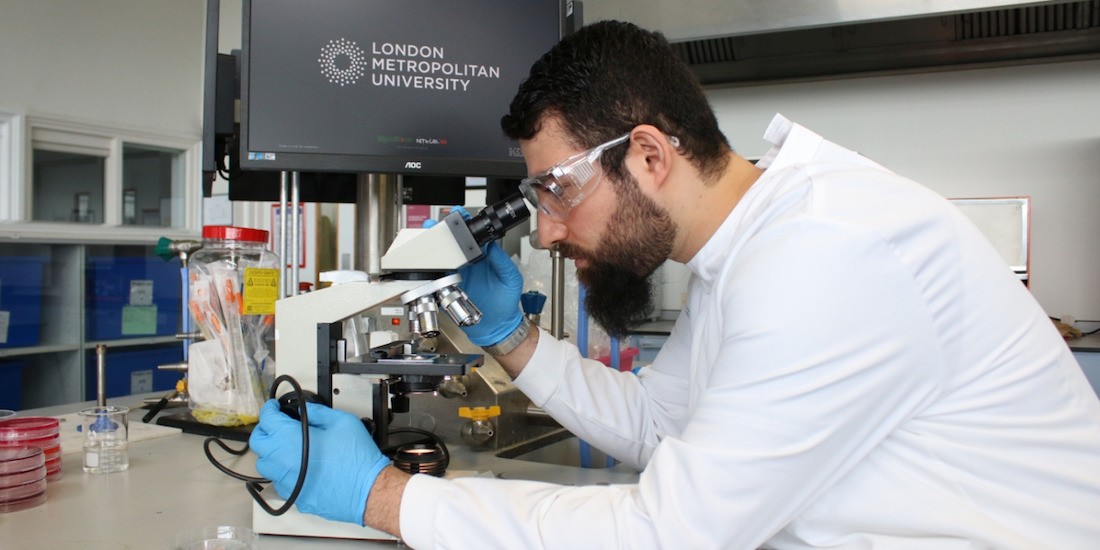 Money under the microscope - Financial comparison website money.co.uk, teamed up with the London Metropolitan University to put money under the microscope to discover what’s really lurking in your wallet - the findings are not for the faint hearted. Dr Paul Matewele, Professor of Microbiology at London Met, and his students took 36 samples from a random selection of all denominations of coins and notes. The microbiologists studied the bacteria in a controlled lab environment over a period of 8 weeks....
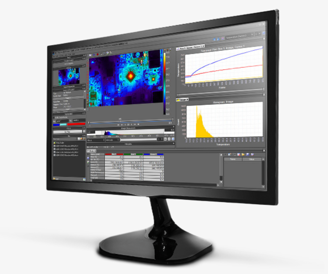 FLIR Systems announces update version 4.40 of its popular thermal measurement, recording and analysis software - ResearchIR. ResearchIR 4.40 is a powerful, yet easy to-use thermal analysis software package for research grade uncooled and cooled cameras from FLIR Systems. The latest revision of this software suite provides highly intuitive advanced camera...
 Phasefocus™ is delighted to announce the launch of a new corporate website. With a fresh look and feel, the new site has been designed to improve the end-user experience, providing simple, logical, navigation, allowing visitors to rapidly locate and access a wealth of product and application information from a single location. Visitors to the site can now browse the wide range of Livecyte™ applications, from where they can...
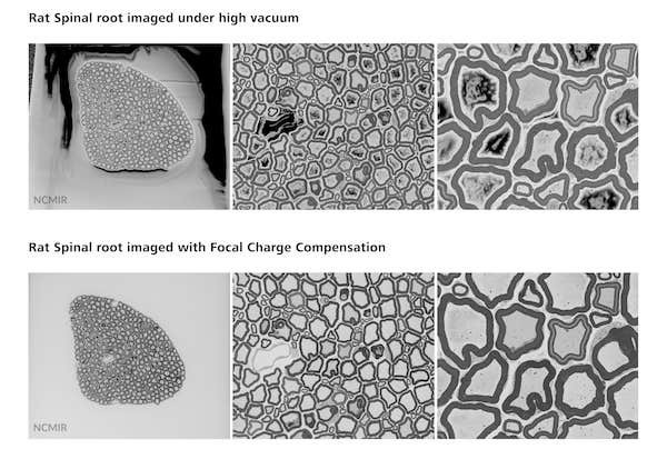 High resolution 3D block face imaging for biological samples with fast acquisition rates and minimal sample damage. In collaboration with the National Center for Microscopy and Imaging Research (NCMIR) at the University of California San Diego, ZEISS releases a new Focal Charge Compensation module for block face imaging with ZEISS GeminiSEM and 3View® from Gatan, Inc. Focal Charge Compensation...
 Keynote speakers have been announced for HSI 2018, the 7th international conference on Hyperspectral Imaging & Applications, co-located with Photonex Europe taking place on 10th & 11th October at the Ricoh Arena, Coventry, UK. HSI 2018 brings together academic and industrial users to share their knowledge of innovations in hyperspectral imaging cameras, technology and applications....
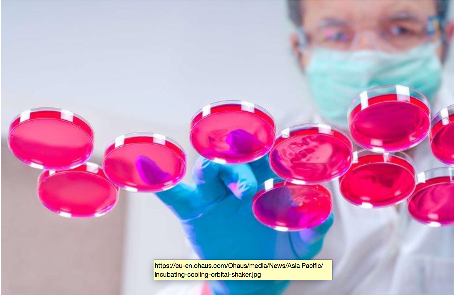 Understanding the process of interaction between healthy and diseased cells provides a better understanding of the life processes of organisms, resulting in more effective medicines, new vaccines. In every successful relationship, communication is key. This can be among friends in a social setting, at home with family or in the workplace with colleagues....
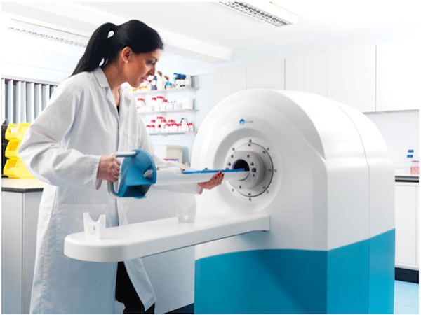 Founder and Chairman, Dr David Taylor, of Guildford based MR Solutions has been shortlisted as an Innovation Entrepreneur of the Year for London and the South East in the 2018 NatWest Great British Entrepreneur Awards. The awards have had a record number of entries with businesses from across the south east vying for a place on the shortlist...
 The University of Glasgow has opened an innovative new structural biology centre, home to a cutting-edge electron microscope – the first of its kind in Scotland – which will be used to image biological molecules at the atomic level. The technology will be used to support vital research into diseases posing the greatest threat to human and animal health, providing greater capabilities in areas such as vaccine development, cancer research, and drug design and discovery....
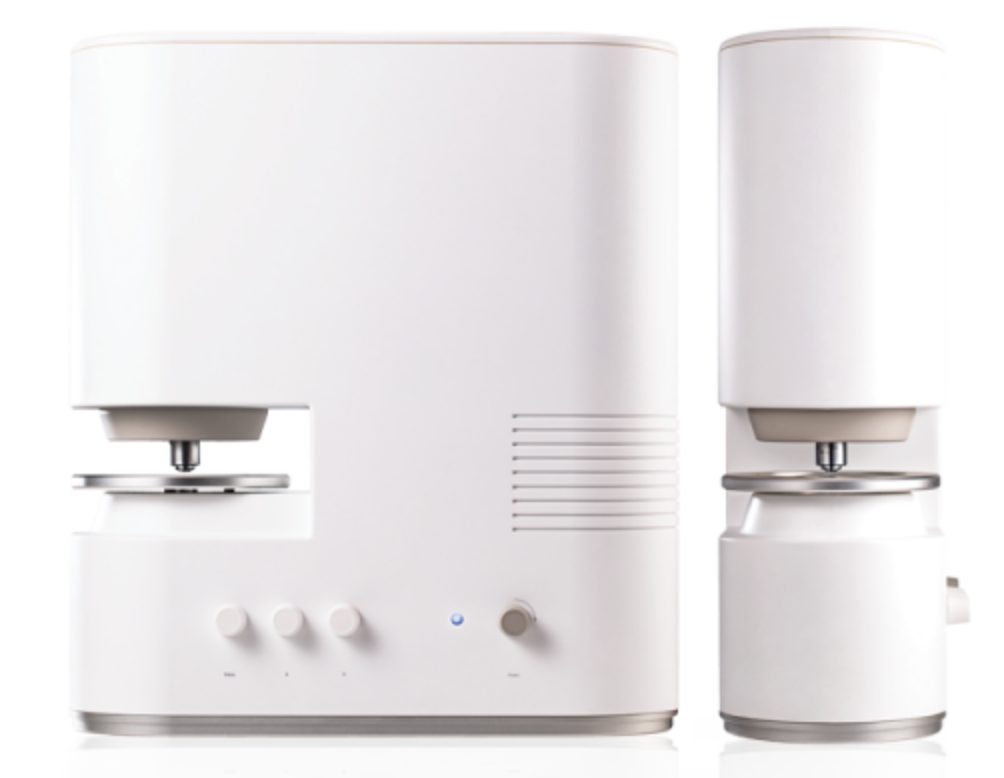 Tomocube’s revolutionary holotomography technique delivers 3D structural and chemical information quickly and simply without any labelling or pre-preparation. A spin-out company from the renowned Korea Advanced Institute of Science and Technology in Daejeon has been established to commercialize the revolutionary Holotomography (HT) microscopy developed...
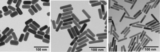 Creative Diagnostics recently launches fluorescent gold nanorods, labeled with various fluorophores for multimodal imaging for the global life science marketplace, which can be applied in various research fields including drug delivery, photothermal therapy, multimodal imaging, dark-field scattering imaging, computed tomography, and so on.
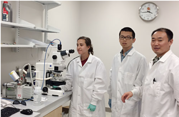 Market leaders in temperature-controlled microscopy and leaders in sample characterization solutions, Linkam Scientific Instruments, reports on the research of Professor Xiaoming “Shawn” He of the Fischell Department of Bioengineering at the University of Maryland. Professor He's research is focused on developing multiscale biomaterials to engineer living cells and tissues for two applications: Cancer treatment and cell-based medicine...
 A team of scientists at the Sainsbury Laboratory Cambridge University (SLCU) have discovered a rare mineral in alpine plants. Vaterite, a form (polymorph) of calcium carbonate, holds enticing potential as a new material for industrial and medical applications. This is the first time that vaterite has been found in plants. The discovery of vaterite was made through a collaboration between SLCU and Cambridge University Botanic Garden....
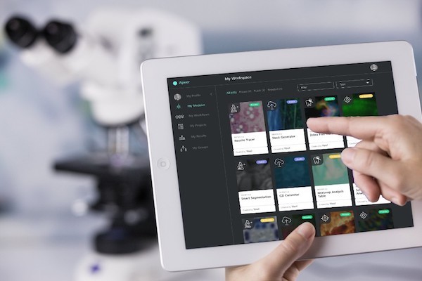 ZEISS presents the initial release of its cloud-based digital microscopy platform, known under the name APEER at the Microscopy & Microanalysis conference (M&M) in Baltimore, USA. Microscopy users will be able to automatically process images in the cloud by leveraging application workflows for 3D reconstructions, staining or segmenting...
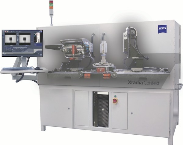 ZEISS expands X-ray imaging portfolio with ZEISS Xradia Context microCT. Built on renowned ZEISS Xradia platform and field-convertible to ZEISS Xradia Versa X-ray microscope. ZEISS introduces a new X-ray imaging instrument, ZEISS Xradia Context microCT, a large field-of-view, non-destructive 3D X-ray microcomputed tomography system. ZEISS Xradia Context is built on time-tested ZEISS Xradia technology, reaping crossover benefits of years of...
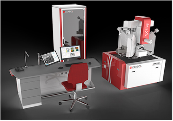 CAMECA, a world leader in scientific instrumentation and metrology solutions, is pleased to announce the launch of SXFive-TACTIS, the newest addition to the CAMECA line of high-end microanalytical instruments, and the only Electron Probe Microanalyzer (EPMA) in the world with a touch-screen interface. “We are very proud to introduce the SXFive-TACTIS EPMA, which represents the latest evolution in our SXFive product line,” comments Sanjay Kamtekar...
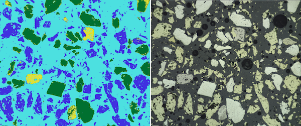 First ZEISS ZEN Intellesis solution enables segmentation of correlative microscopy datasets.
 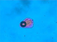 Linkam Scientific Instruments, reports on the research of Dr Jamie Wilkinson of the Natural History Museum in London where he uses temperature-controlled microscopy to characterize mineral deposits. Dr Jamie Wilkinson is a Research Leader in Mineral Deposits in the Department of Earth Sciences at the Natural History Museum, London. He is a member of the London Centre for Ore Deposits and Exploration (LODE) Research Group...
 JPK Instruments, a world-leading manufacturer of nanoanalytic instrumentation for research in life sciences and soft matter, reports on how the Nano Imaging Lab of the Swiss Nanoscience Institute uses JPK’s versatile NanoWizard® 4 AFM system. Dr Monica Schönenberger has developed a flexible, professional service unit for atomic force microscopy. As a member of the Swiss Nanoscience Institute (SNI)...
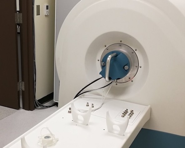 MR Solutions has installed Hawaii’s first preclinical cryogen-free MRI imaging system to help researchers improve the rate of kidney disease detection through research. This is very good news for Hawaii which has a very high incidence of kidney disease - one in seven residents on the island has chronic kidney disease....