
Channels
Special Offers & Promotions
Olympus scanR 3.1
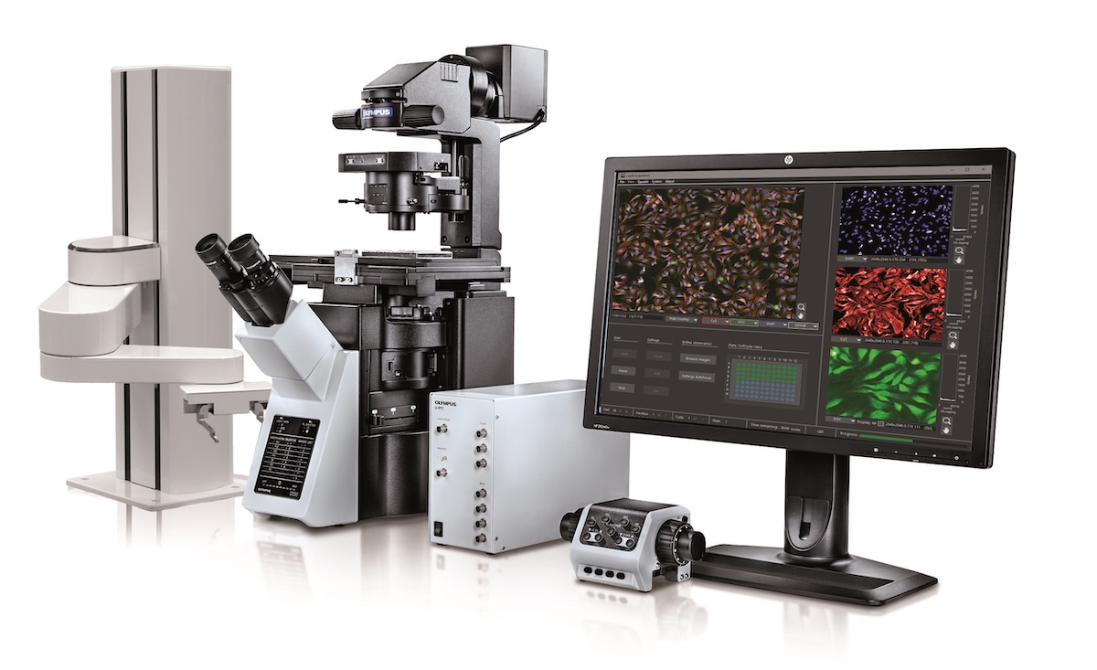
The Olympus scanR 3.1 high-content screening station uses the power of AI to carry out groundbreaking analyses of live cells – analyses that until recently seemed impossible. Its software’s accuracy, robustness and ease of use improve both data quality and cell viability, enabling fast analyses and confidence in results.
Olympus’ newly launched scanR 3.1 high-content screening (HCS) station fully embraces the capabilities of artificial intelligence (AI) to enable cutting-edge life science research. It combines the modularity and flexibility of a microscope-based setup with the automation, speed, throughput and reproducibility of HCS applications. Using the ‘self-learning microscopy’ concept, scanR 3.1 makes it easy to gather data quickly from large live cell populations for reliable, well-supported experimental results.
To minimize setup time, the self-learning approach makes use of a short, one-time training phase in which the software uses a quickly acquired set of images to generate ‘ground truth’ data without requiring human annotations. It then uses convolutional neural networks to autonomously create robust algorithms that can rapidly analyze large sets of images.
One application that clearly shows the power of AI in HCS is label-free quantification of live cells. Olympus’ scanR HCS software can reliably derive nuclei positions in micro wells solely from brightfield transmission images – with an accuracy that rivals fluorescence. Quantifying live cells from brightfield images instead of fluorescence shortens exposure times, avoids using genetic modifications or nucleus markers, and saves fluorescence channels for other markers. These benefits reduce phototoxicity and lead to simpler, faster image acquisition and better cell viability.
Reliable low light analysis has also become possible thanks to scanR’s AI-based imaging software. Olympus has shown that its software accurately detects DAPI-labeled cells at only 0.2% of the optimal light intensity. It can even distinguish different stages of the cell cycle, based on the intensity of the signal, providing better insights and improving reproducibility.
Dr. Manoel Veiga, application specialist at Olympus Soft Imaging Solutions, commented on the launch, saying: “This exciting new scanR system is good news for a whole range of life science applications. Low light and label-free analysis are only examples of how automated detection using an AI-based approach can improve the accuracy and reproducibility of HCS. Olympus’ self-learning microscopy approach for scanR 3.1 combines these benefits with a user-friendly workflow that requires little human interaction during the training stage. This makes it easy for life science researchers to harness the power of HCS data while benefitting from a fast, simple setup.”
News Channels
- Latest News
- New Laboratory Products
- Industry News
- Laboratory Automation | IT Solutions
- Microscopy | Image Analysis
- Separation Science
- Research | Case Studies
- Video Presentations
- Events | Conferences
Subscribe to any of our newsletters for the latest on new laboratory products, industry news, case studies and much more!
Popular this Month
Top 10 most popular articles this month
Today's Picks
Looking for a Supplier?
Search by company or by product
Please note Lab Bulletin does not sell, supply any of the products featured on this website. If you have an enquiry, please use the contact form below the article or company profile and we will send your request to the supplier so that they can contact you directly.
Lab Bulletin is published by newleaf marketing communications ltd.
Media Partners