
Channels
Special Offers & Promotions
Olympus Europa Holding GmbH
Products
Contact Olympus Europa Holding GmbH
All articles from Olympus Europa Holding GmbH
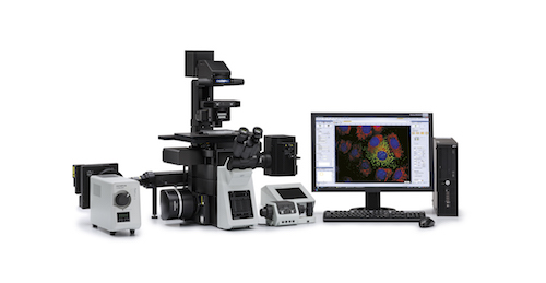
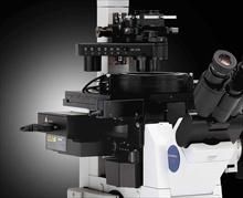
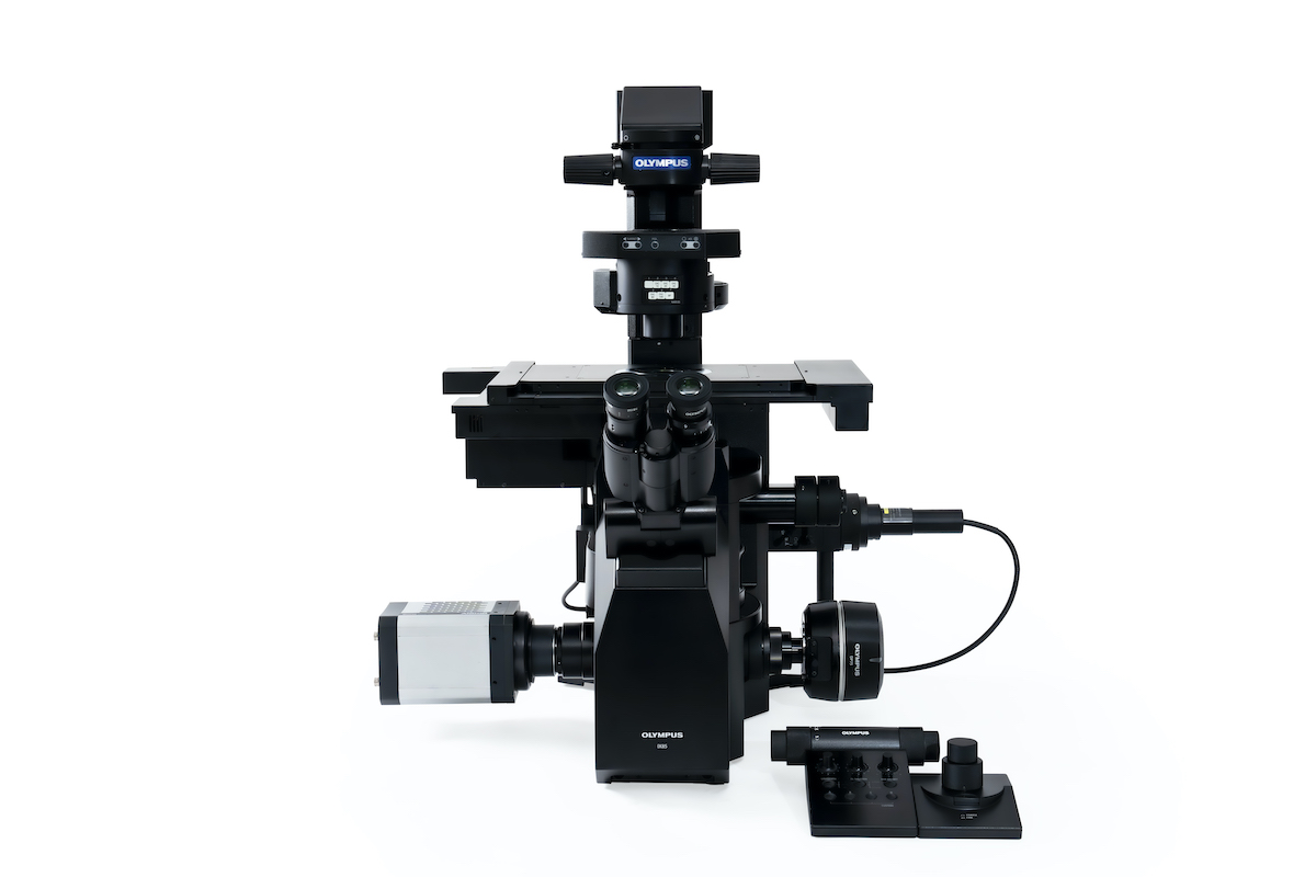 Evident recently announced the release of its IXplore™ IX85 automated inverted microscope system. With an industry-leading 26.5mm field number (FN), combined with new advanced imaging tools that ensure clear, accurate results, this new system empowers researchers to capture more data and uncover new insights faster than ever before. It also delivers an unmatched level of customizability, allowing users to design or build an intelligent, high-performance imaging platform that meets their specific goals...
 Beauty of cosmic proportions found in microscopic imaging. Evident unveiled the winners of its 5th annual Image of the Year contest, an awards competition that recognizes the world’s best in scientific microscopic imaging. The winners were selected from submissions from 29 countries around the world. For the first time, the competition included a video category to showcase the art of capturing small changes in motion under the microscope...
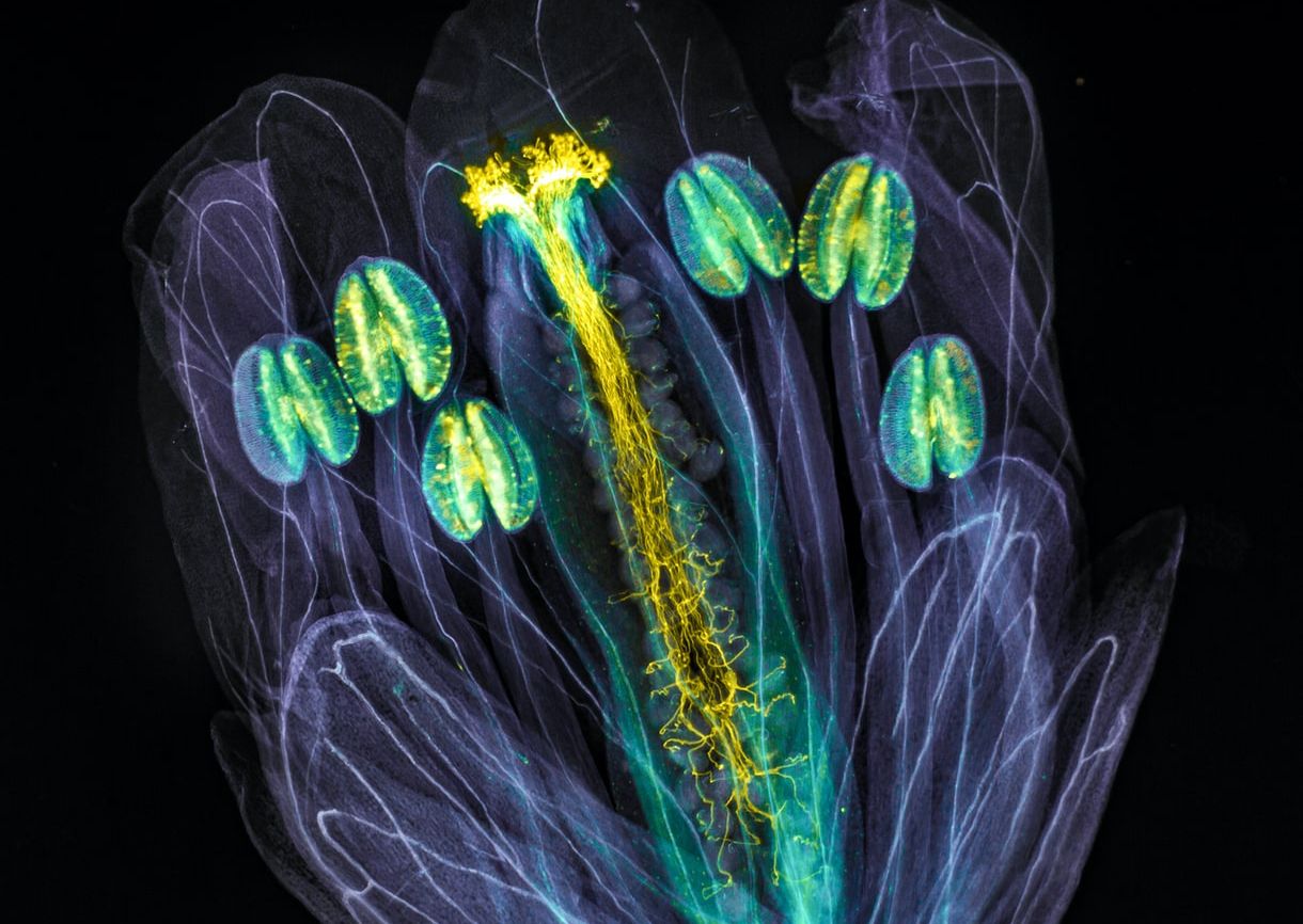
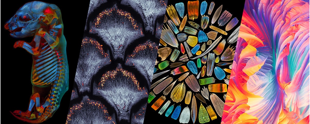
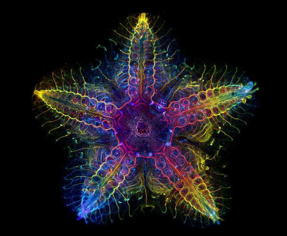 Evident unveiled the winners of its fourth Global Image of the Year Scientific Light Microscopy Award, an annual competition that recognizes the best in scientific imaging worldwide. The winners were selected from 640 images submitted from 38 countries around the world. Laurent Formery from the United States was selected as the global winner for his stunning image of the nervous system of a juvenile sea star (Patiria miniata)...
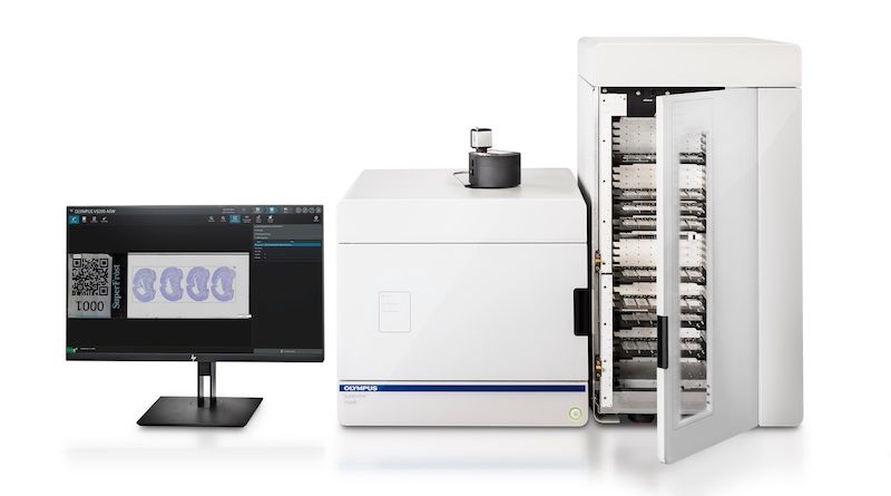
The SLIDEVIEW™ DX Combines Microscope-Quality Images with Fast Slide Scanning for Efficient Digital Pathology
Jun 14, 2023
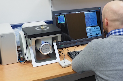
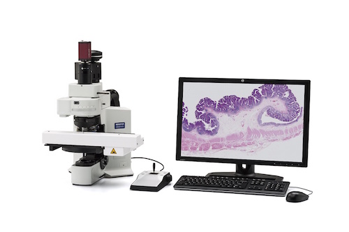
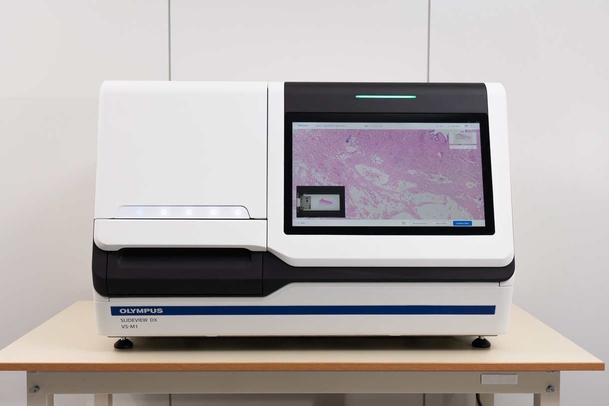 The new Evident SLIDEVIEW™ DX VS-M1 whole slide imaging system is a digital pathology solution that delivers high-quality slide images at high speed, helping pathologists make diagnoses quickly and efficiently. Built with renowned optics and advanced digital technology, the SLIDEVIEW DX slide scanner is a complete digital pathology solution. The SLIDEVIEW DX scanner provides microscope-quality images onscreen...
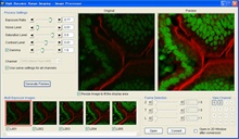
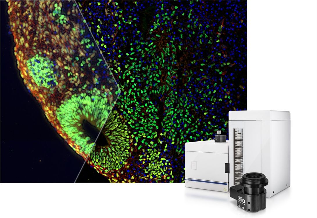 Get Better Images from Deep within Your Sample. The new SLIDEVIEW™ VS200 v. 4.1 software supports a host of new features, including the SILA optical sectioning device that enables you to obtain high-contrast images from deep within your sample. The SILA optical sectioning device uses speckle illumination combined with HiLo microscopy to achieve high-contrast images. This camera-based technology quickly captures two illuminated images that are then mathematically processed to remove out-of-focus light...
 Building on the momentum from last year’s conference, the 2023 Evident Organoid Conference will highlight organoid innovations that are poised to accelerate progress in drug research and discovery. Taking place on February 8, 2023, this virtual conference brings together experts from the Asia-Pacific region to discuss their latest research discoveries...
 Evident announced the opening of its new Asia-Pacific (APAC) headquarters in Singapore, marking its ongoing commitment to expand its business and operations in the APAC region. The headquarters leads Evident’s strategy, technology and innovation in the APAC region, which includes operations in Australia, New Zealand, India and Korea. The company has doubled its headcount in Singapore since the beginning of 2022 and plans to further expand its presence...
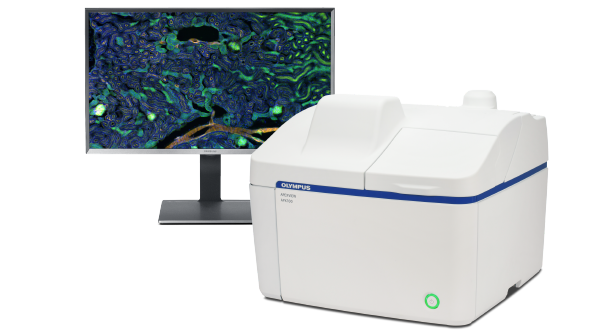 Do you want publication-quality, multidimensional microscope images without the complexity of typical high-end technology platforms? Then discover the APX100 digital imaging system from Evident—a user-friendly, all-in-one microscope that will take your images to the next level in just a few clicks. The APX100 is packed full of features that makes collecting data and acquiring expert-quality images simple and efficient. Find out more!...
 Evident, a wholly owned subsidiary of Olympus Corporation, announced its fourth Global Image of the Year Scientific Light Microscopy Award is now open for entries through Feb. 28, 2023. Each year, the competition recognizes the best in scientific imaging worldwide. For the first time, the contest welcomes materials science images in addition to life science images to show the versatility of the art of science...
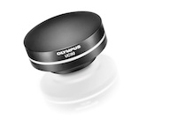
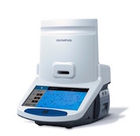
 Evident, a wholly owned subsidiary of Olympus Corporation, is proud to announce that its Olympus Provi™ CM20 incubation monitoring system is the 2022 recipient of the prestigious "Best of the Best" award in the Product Design category of the Red Dot Design Award. This Award is bestowed on a product that the Red Dot Jury deems as groundbreaking and is the highest honor given in this world-renowned design competition...
 Evident, a new wholly owned subsidiary of Olympus comprised of its former Life Science and Industrial divisions, unveiled the winners of its third Global Image of the Year Life Science Light Microscopy Award, an annual competition that recognizes the best in life science imaging. The winners were selected from nearly 800 images submitted from 49 countries around the world...
 Olympus Corporation (“Olympus”) announced the completion of the separation of its Scientific Solutions business to a wholly-owned subsidiary, Evident Corporation (“Evident”). Following the divestiture of its Imaging business, this move further signifies the company’s long-term strategy to cement its position as a major player in the medtech industry...
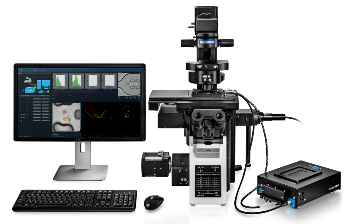 Olympus, a leading global manufacturer of optical and digital precision technology, announced a partnership with evorion biotechnologies, a pioneer in advanced single-cell analysis using unique microfluidic systems. By merging evorion’s cutting-edge 3D hydrogel bead technology and Olympus’ leading microscope systems, the collaboration introduces a seamless and largely automated workflow solution to capture functional phenotypes of individual cells´ behavior over time...
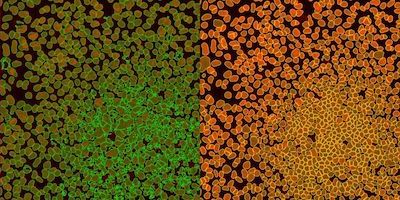 The scanR high-content screening (HCS) station provides fully automated image acquisition and data analysis. Version 3.3 improves the deep-learning technology’s capabilities to reliably separate objects in biological samples using instance segmentation, the ability to detect and delineate distinct objects of interest in an image. Using a self-learning microscopy approach, the scanR system’s AI automatically analyzes data in an assay-based workflow. The deep-learning technology can detect cells, nuclei and subcellular objects...
Olympus Announces First Results of Its AI-Based Pathology Diagnostic Tool for Gastric Cancer
Nov 29, 2021
 Olympus’ third annual Global Image of the Year Life Science Light Microscopy Award is now open for entries through Jan. 31, 2022. Each year, the competition recognizes the best in life science imaging worldwide to inspire and showcase art through microscopy. Contestants may enter by uploading up to three images, with a description of the equipment used, at the website. Winners will be selected by a jury and announced in April 2022.
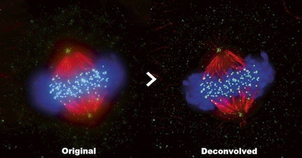
 After a successful first summit in April 2021 that concentrated on moving forward in a post-pandemic world, the second Olympus Discovery Summit will focus on how microscopy imaging can advance research. Presentation topics include the use of complementary imaging technologies to understand the immune system, deconvolution techniques to improve image quality, 3D imaging, how AI is helping researchers analyze large amounts of microscopy image data and more...
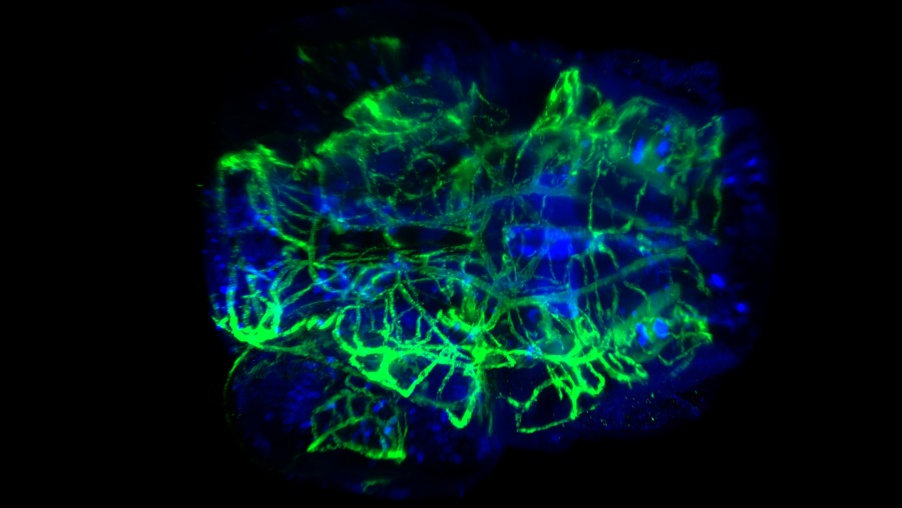
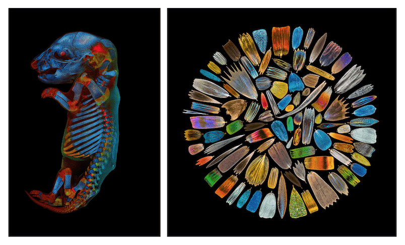 Olympus unveiled the winners of their second Global Image of the Year Life Science Light Microscopy Award, an annual competition that recognizes the best in life science imaging. The winning images were selected from a record number of entries—nearly 700 submissions from 61 countries. Global Winner, Werner Zuschratter from Germany was selected as the global winner for his eye-catching image of a whole rat embryo captured with a confocal microscope....
 Olympus’ next virtual life science event, the Olympus Discovery Summit—Looking Forward: A New Era of Research will take place on April 27–29, 2021. The Olympus Life Science team has organized this opportunity for the microscopy community to virtually share its expertise and learn about important advances in technology. During this free three-day summit, attendees can enjoy a full schedule of customer presentations, tech talks, roundtable discussions and product demos....
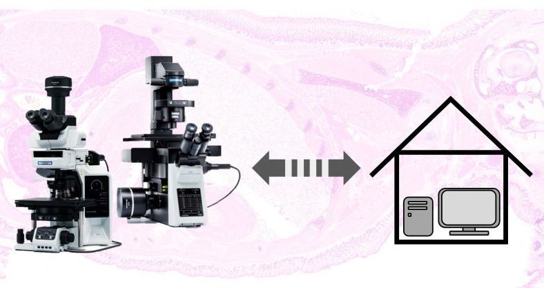 As more lab personnel work from home to practice social distancing, employees are using remote work IT platforms to communicate and stay productive. But this change in the way scientists and lab personnel work means labs must adapt their methods to fully leverage remote platforms. If you’re not sure where to start, this blog can help. Here are six tips to help you successfully implement remote microscopy...
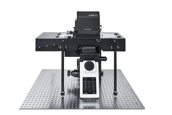 Olympus, a leading manufacturer of high-end research microscopes, and Cytosurge, a precision manufacturer of cell manipulation technologies, have entered a co-marketing agreement to become a complete system provider to the scientific community’s growing need for next-generation single-cell and CRISPR genetic manipulation solutions....
 To capture the importance of this initiative, and in anticipation of the grand opening of the Ellison Institute’s new building, Olympus has created a video, highlighting the relationship between imaging analysis tools and precision cancer medicine. One focus of the partnership is encouraging projects that engage translational oncology and precision anti-cancer drug screening...
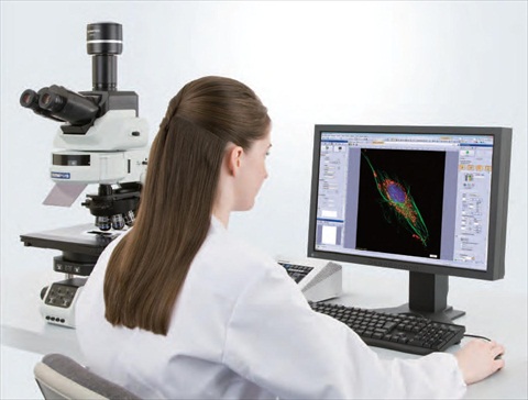
 Olympus cellSens imaging software improves the efficiency of your research with accurate object detection and segmentation. Leveraging the power of deep learning, Olympus cellSens imaging software for microscopy offers significantly improved segmentation analysis, such as label-free nucleus detection and cell counting, for more accurate data and efficient experiments....
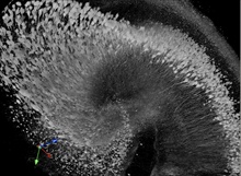
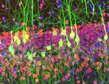 Olympus is proud to announce the winners of its first Global Image of the Year Life Science Light Microscopy Award, a competition that recognizes the best in life science imaging worldwide. Ainara Pintor from Spain was selected as the global winner for her vibrant image of an immunostained mouse brain slice with two fluorophores. She named her winning image “Neurogarden” because it reflects the brain’s complexity....
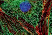
 We live in a three-dimensional ever-changing world. Yet in microscopy, our ability to image in 3D and at the speeds required to observe fast living processes remains a challenge. Compared to other light microscopy techniques, fluorescence microscopy can achieve high specificity and contrast, though there are still major limitations in signal-to-noise ratio and phototoxicity.
Olympus SLIDEVIEW VS200 Solution with Reliable, Flexible, and High-Throughput Slide Scanning
Nov 12, 2019
 The new Olympus SLIDEVIEW VS200 slide scanner captures high-quality virtual slide images and offers flexibility to empower advanced quantitative image analysis for brain, cancer and stem cell research, as well as drug discovery. Simple to use, its intuitive workflows enable users to start scanning a slide in as few as two clicks. By employing Olympus X Line objectives at the core of the system, users obtain flatter images with a wider field of view....
 At this year’s European Congress of Pathology (ECP), Olympus will demonstrate how its 100 years of optical experience and the latest in ergonomic microscope design work together to make pathologists’ lives easier. Olympus will be displaying the culmination of 100 years of expertise in optics at booth R43 of the ECP in Nice from 7 to 11 September 2019. The 4K UHD UC90 camera, the new high-performance X Line objectives and the ergonomic BX46 microscope have each been carefully designed to enhance pathology workflows.
 Olympus’ Image of the Year Award recognizes the best in life science imaging worldwide. Participants can win a CX43 microscope with a DP27 digital camera, X Line objectives, or an OM-D E-M5 Mark II camera. Those interested in participating can enter until 31 January 2020 by uploading images at the website. Winners will be selected by a jury panel and announced in March 2020....
 The Olympus scanR 3.1 high-content screening station uses the power of AI to carry out groundbreaking analyses of live cells – analyses that until recently seemed impossible. Its software’s accuracy, robustness and ease of use improve both data quality and cell viability, enabling fast analyses and confidence in results. Olympus’ newly launched scanR 3.1 high-content screening (HCS) station fully embraces the capabilities of artificial intelligence (AI) to enable cutting-edge life science research....
 Today’s educational landscape is changing rapidly thanks to technological advances. Introducing wireless capabilities to science classrooms enables lecturers to teach and students to learn in an engaging digital environment that promotes teamwork. Equipped with a WLAN-capable Olympus EP50 camera, every microscope in a classroom becomes a wireless imaging system...
Olympus Europe and Cytosurge Partner to Enhance Pharma Drug Development and Single Cell Research
Jul 19, 2019
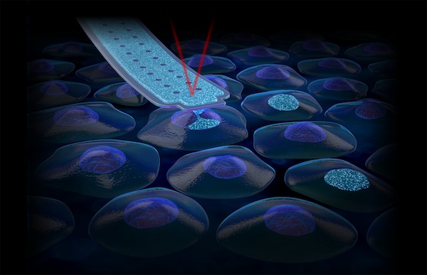 Olympus Europe and Cytosurge have partnered in order to accelerate common innovation of next-generation tools for single cell experiments and to expand the distribution of the versatile FluidFM BOT single cell research tool. The ultimate goal of the new co-marketing partnership is to fight diseases and improve human health by enhancing drug development research in pharma, revolutionize single cell gene editing...
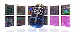
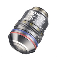
 Olympus delivers a breakthrough to life science microscopy with the launch of its next-generation X Line objectives. Ultra-thin lenses manufactured by a revolutionary polishing technique enable the objectives to simultaneously boost numerical aperture, flatness and chromatic correction – providing exceptional image quality for researchers and clinicians alike. As a leader in optics for 100 years, Olympus has broken down barriers to imaging quality....
.jpg) The Olympus high-sensitivity SpinSR10 super resolution microscope combines fast 0.005 seconds/frame acquisition speed and 120-nanometer super resolution with improvements that provide clearer images and three times the brightness of the standard SpinSR10 model. Super resolution microscopes are used in medical and biological research to observe the internal structure of cells in fine detail, and brightness and clarity are essential to image quality....
The Olympus high-sensitivity SpinSR10 super resolution microscope combines fast 0.005 seconds/frame acquisition speed and 120-nanometer super resolution with improvements that provide clearer images and three times the brightness of the standard SpinSR10 model. Super resolution microscopes are used in medical and biological research to observe the internal structure of cells in fine detail, and brightness and clarity are essential to image quality....
 Helping drive research forward at a faster rate, Olympus cellSens 2.1 speeds up image analysis with improved capabilities for flexible camera and microscope control. The updates ensure cellSens remains a powerful tool covering a wide range of applications and benefitting routine microscopy users as well as advanced imaging facilities....
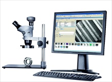
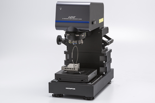 Saving time with a four times higher scan speed, Olympus’ new confocal microscope improves efficiency in industrial inspection.
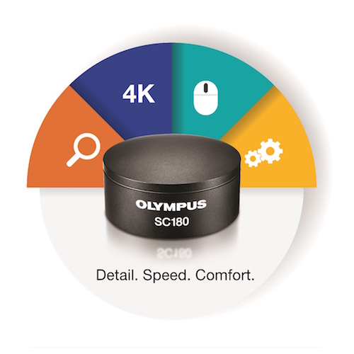 With unprecedented detail, especially at low magnifications, Olympus’ 18-megapixel SC180 camera facilitates precise sample documentation and audience engagement alike.
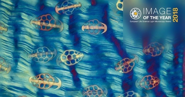
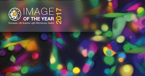 For the chance to win a microscope, applicants can simply submit their best life science light microscopy images taken with Olympus equipment to the campaign website. Olympus’ Image of the Year European Life Science Light Microscopy Award recognizes the very best in life science imaging....
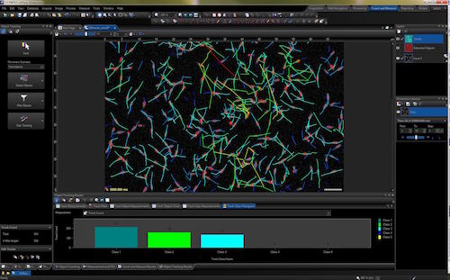 Olympus has released version 1.17 of its life science imaging software cellSens. The new cellSens features an automatic object tracking function – a dedicated solution to analyse and document dynamic processes within living samples....
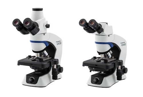 With a maintenance- and centring-free LED light source and a range of ergonomic features, the CX43 and CX33 are ideal for high-throughput, regular use. Combining exceptional user comfort with bright, uniform illumination, Olympus has launched two new microscopes optimised for long periods of routine observation....
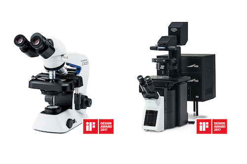 Globally renowned as a symbol of design excellence, the iF design awards celebrate the best in user-focused, ergonomic and efficient design. With over 5,000 submissions from 70 countries, Olympus Scientific Solutions Division has received two of these prestigious awards for its CX23 upright microscope and FLUOVIEW FV3000 confocal laser scanning...
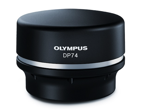 With a 60 frames per second live image and a 20.7 megapixel resolution it delivers a lifelike experience on-screen while also introducing intelligent features to make everyday operation faster and more convenient. Helping to save time and increase comfort at the microscope, Olympus’ new DP74 camera for brightfield and fluorescence delivers intelligent imaging....
 Accurate cell confluency measurements are now quick to perform on cell cultures with the Olympus CKX-CCSW confluency checker software. Without having to remove cells from the vessel, the software quantifies the exact growth density to quickly help scientists decide on the next step and optimise cultivation conditions. The new software complements the Olympus CKX53 cell culture microscope and Cell Counter...
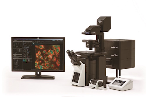 Built for fast, stable and accurate measurements of biological reactions within living cells and tissues, Olympus’ FV3000 offers flexibility for all live-cell imaging applications, providing high-resolution images of structures and dynamic intracellular processes. Succeeding the well-established FV1200, the FV3000 is controlled via a new, intuitive software interface so even novice users can generate high-quality data and images...
 Unravelling the mechanical properties of bone with micro-indentation testing has significance in both health and disease. At the University Medical Center Hamburg-Eppendorf, Dr. Björn Busse and his research group have advanced their micro-indentation testing technique using an
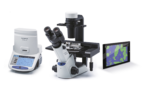
 Streamlining the cell culture workflow in a range of application areas, Olympus’ renowned optical expertise has given rise to the new
 These new technologies deliver fast and accurate multi well time-lapse acquisition, stable focus during long time-lapse studies and an easier user interface. Taking well plate imaging to the next level, Olympus presents two innovative tools for the customisable IX3 inverted microscopes. The new IX3-ZDC2 module offers cutting-edge Olympus focal drift compensation technology, enabling accurate focus stability during long...
 Maximising the information captured from life and materials science samples is now possible with ease and simplicity, with the new Olympus UC90 microscope camera. Thanks to a range of innovative features, the UC90 excels in capturing and documenting real-life details, while a 4K UHD mode supports scientists in overcoming the limitations of the oculars with the many benefits of complete on-screen operation. In-depth sample...
 The Cell Counter model R1 is a cost-effective solution that saves time and simplifies the cell counting process. Engineered with leading-edge technology in a portable design, the new Olympus Cell Counter model R1 offers user-friendly and cost-effective cell counting for routine cell culturing. Accurate cell counts are important for a variety of research applications, including regenerative and drug medicine. Rather than relying on a human operator to manually count...
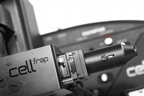 Enabling the easy addition of photomanipulation techniques to imaging platforms, Olympus has launched a new cellFRAP deck system for the popular IX3 ‘open source concept’ IX83 and IX73 microscopes. Olympus cellFRAP is designed with researchers in mind, providing highly accurate and flexible live-cell photomanipulation with various evaluation options for data presentation needs. Unlike conventional widefield-based FRAP systems, Olympus’ cellFRAP...
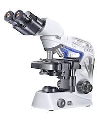 Every aspect of use has been taken into account during the design process providing an effortless user experience across all workflows. Enabling maximum comfort and ease-of-use for routine observations, the high-performing, ergonomically-designed, compact and robust CX23 microscope is packed full of well-considered, clever features to enhance every step of the workflow. Delivering Olympus’ renowned high-quality optics and featuring updates to increase portability...
 High resolution scanning is delivered via a range of features including a new scanning stage and support for a sensitive sCMOS camera, tailored to the needs of fluorescence scanning applications. Olympus’ award winning VS120 virtual slide systems have been updated to enable precision and accuracy in high-speed fluorescence and brightfield scanning – with the ability to scan a brightfield slide in under two minutes. The product range...
FluoView FVMPE-RS multiphoton system - two new microscopes for live cell and in-vivo imaging
Feb 19, 2015
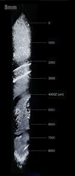
 The flagship offering in the range of Olympus biological microscopes, the FluoView FVMPE-RS series is widely used in life science research. Its high-speed scanner allows observation of ultra-rapid biological responses, and the system can obtain vivid images from as deep as 8 mm below the tissue surface....
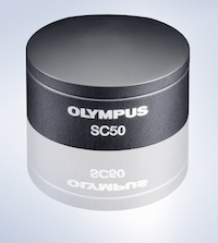 Designed for the convenience of everyday use and ideal under low light conditions, the new 5 megapixel SC50 microscope camera from Olympus is built on the latest CMOS chip technology to deliver improved speed, sensitivity and resolution at a truly affordable price. Achieving Full HD images at 32 fps, high speed imaging with the SC50 captures and records dynamic events. The camera delivers clear, high quality images to nearly every kind of...
 Discovering and understanding novel strategies for historic conservation, Dr Olivier Schalm’s research at the Department of Conservation Studies, University of Antwerp (Belgium), has benefited from the latest in high-resolution light microscopy with the Olympus DSX500 Opto-digital system. Introducing cutting-edge digital imaging technologies and retaining true colour information at magnifications of up to 4,000x, this approach has even...


 With its high speed and precision performance, the FVMPE-RS is designed for electrophysiology and optogenetics studies. It is also a good match for applications such as high-speed calcium and in vivo imaging, peristalsis and blood flow studies, mosaic imaging, connectomics and functional brain imaging, stem cell research, and any field that requires precise colocalisation, uncaging, simultaneous imaging/stimulation, extensive real-time signal processing, or multipoint mapping....
 Demonstrating the very latest in live cell imaging technologies, Olympus will exhibit its range of IX3 inverted microscope frames with associated software and accessories at the upcoming European Molecular Biology Organisation (EMBO) Meeting 2013, taking place in Amsterdam on 21-24 September. Delegates of the “Seeing is Believing –Imaging the Processes of Life” Symposium hosted by EMBO in collaboration with the European Molecular Biology Laboratory (EMBL), which takes place on the 3-6 October in Heidelberg (Germany), will also have the chance to discover how the Olympus open source microscopy concept is advancing live cell imaging...
 Enhancing the workflow of research and diagnostic centres alike, the latest range of Olympus pathology microscope systems and accessories will be on show at the 25th European Congress of Pathology (ECP) 2013, 31 August – 4 September in Lisbon, Portugal. Delegates can visit booth 14 to view the Olympus BX3 range of upright microscopes and the Olympus VS120 virtual slide scanner...
 The IX3 series can now be further optimised for a wider range of live cell imaging applications including in vitro fertilisation, with motorised functionality for accuracy and easy operation. Extending the capabilities of the fully customisable IX3 inverted microscope frame, Olympus has expanded its range of compatible, interchangeable optical modules. Ease-of-use and accuracy are vastly improved via new motorised components, which also present a cost-efficient means to gradually upgrade to a fully motorised microscope system....

 Focusing on ease of use for routine applications, the latest cellSens software release, version 1.8, offers flexible and user-centric imaging, processing, analysing and reporting. New and enhanced functions include a fully customisable user interface, precise frame-by-frame sample navigation and easy one-button switching between wells. For a wide range of life science applications, cellSens provides easy control of the workflow. The software binds together Olympus frames, cameras and high-quality optics to form complete, highly precise and user-friendly microscopy systems....
 The scan^R 2.4 high content screening station is the latest addition to the growing collection of microscopy systems based upon the new IX3 high-end inverted microscope frame. Thanks to its unique “open source” design concept, the IX3 is offering a completely modular and flexible approach to live cell imaging. Built around a swappable deck design, optical modules can be easily exchanged into the accessible infinite light path, moulding the IX3 microscope to the diverse requirements of the user. The updated Olympus scan^R 2.4 high content screening station can now be incorporated with the most advanced model of the IX3 range, the fully automated IX83, combining benefits of a modular frame design with increased scan speed...
 Olympus will exhibit their IX3 inverted microscope systems for live cell applications in booth 46-48 at the Focus on Microscopy 2013 (FOM) annual conference in Maastricht, Netherlands, 24th-27th of March. The new open source frame concept has been designed around an infinite light path made easily accessible with interchangeable optical modules that can be swapped in and out of the light path, enabling the creation of various distinct systems on one inverted microscope frame...

 Olympus has launched the DP80, a unique microscopy camera for both colour documentation and fluorescent detection. Combining a colour and monochrome chip within the same housing, the DP80 provides high resolution brightfield imaging alongside sensitive photon detection. With versatile functionality, the control software permits rapid and automatic exchange between the chips, without switching camera or optical path...

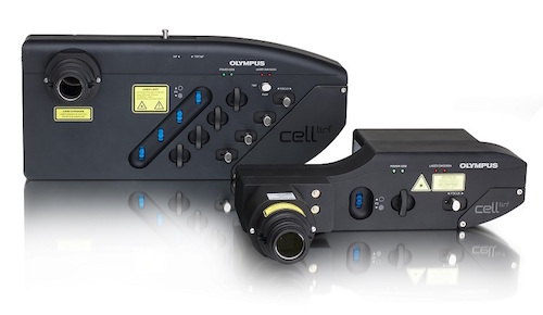
 The new system uniquely combines the accuracy of the newly engineered IX83 frame with enhanced fluorescence sensitivity and simultaneous laser stimulation of cells, making it ideal for advanced life science applications such as FRAP, FLIP and photo-activation. In particular, the new highly-reflective, silver-coated galvanometer scanning mirrors and dual channel GaAsP FluoView PMT module both act to maximise light transfer and detection. This allows for reduced laser power, protecting against the effects of photobleaching and phototoxicity...
 As part of its promise to provide world-class service, Olympus has enhanced its website to guarantee comprehensive, accessible and tailored support for all customers. Based on extensive market research and customer feedback, Olympus completely redesigned its website for life science microscopy. It now offers customers an interactive resource, enabling them to navigate in an application-focused manner towards the products and services that are best suited for their individual requirements...
 The advanced technology of the VS120 creates a “virtual slide”, a high resolution image of the complete specimen that can be electronically stored on a central server for simultaneous viewing anywhere in the world, at a range of magnifications. Awards were obtained in the fields of Colour and Precision, Fluorescence 20X, and Fluorescence 40X. Exhibiting unsurpassed performance in fluorescence microscopy, combined with...
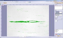
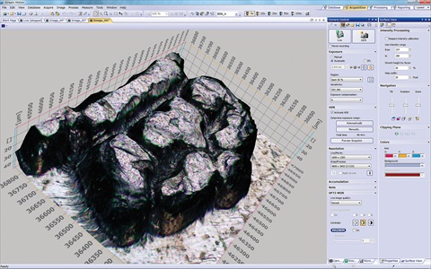 Olympus today launched the latest version of its highly successful OLYMPUS Stream materials science microscopy imaging software family. OLYMPUS Stream 1.8 provides new and improved features, enhancing the overall capabilities of the software to help users fully integrate and automate their workflows. Users will benefit from the advanced automation of a multiple stage location engine, extended data management, new measurement options and new additions to the optional Materials Solutions: Particle Distribution, Porosity, Throwing Power and Phase Analysis. These Materials Solutions enable users to build a fully guided, or even automated, system to precisely match the needs of their materialographic analysis...
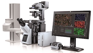
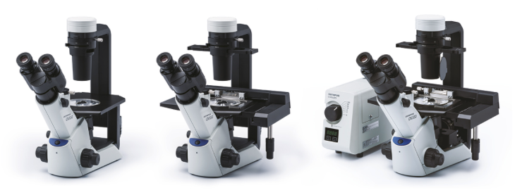
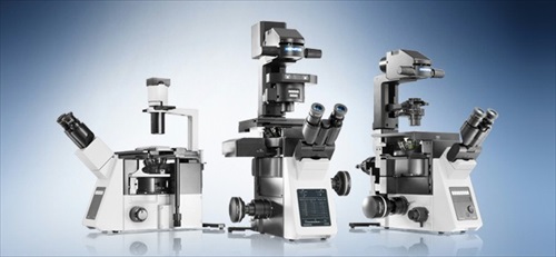
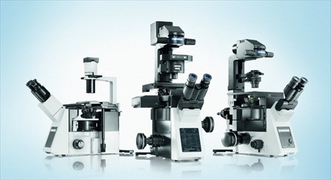 Olympus has released the innovative new IX3 series of inverted research microscope systems for effortless, intuitive live cell imaging and clinical analysis. This includes the fully automated IX83 for high-end research applications, the flexible IX73, which can be configured in manual, semi-motorised or motorised modes, and the easy-to-use IX53 with fluorescent capabilities, which is optimised for the routine examination of tissue samples. Built using worldwide customer feedback and designed to meet the needs of a wide range of users, the new systems offer exceptional ease-of-use and unprecedented optical flexibility via a new, customisable light path...
 Olympus has announced a new release of its powerful and flexible cellSens software (now version 1.7), offering a range of new benefits that allow any user to generate expert results, regardless of experience level. These include the brand new ‘Well Navigator’ Solution, which opens up a new world of flexible, multi-well imaging experiments. cellSens 1.7 also offers High Dynamic Range (HDR) imaging for the perfect exposure of challenging samples, 2D deconvolution for maximising image clarity and a new database model for streamlining the management of large data repositories with many users...
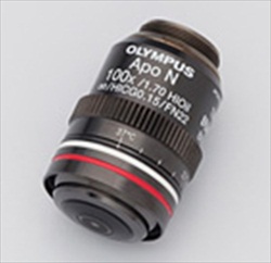 Olympus, a leading manufacturer of professional opto-digital products, has launched the NA 1.7 APON100×HOTIRF objective for the imaging of intracellular molecules. With a world record-breaking numerical aperture (NA) of 1.7, the objective achieves the highest Z-resolution ever recorded for a Total Internal Reflection Fluorescence (TIRF) microscope. The benefits for research using TIRF microscopy include extremely thin penetration depth and high signal-to-noise ratio, making the lens ideal for single molecule dynamic studies and super resolution applications...
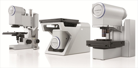 Olympus has today launched its new family of opto-digital microscopes; the DSX series. Through the combination of advanced optics, electronics and Olympus's renowned expertise, the DSX microscopes provide users with superb operating simplicity, and absolute performance reliability. Consisting of three models: the DSX100 free-angle wide zoom microscope, the DSX500 high resolution upright microscope and the DSX500i high resolution inverted microscope, Olympus can provide a complete inspection system to meet the needs of applications across R&D and quality control. These include inspection and analysis within the automotive industry, palaeontology, geology, PCB manufacture, fibre structure, micro-channels, metallography and the geometry of small parts...
 Olympus has introduced the new XLSLPLN25XSVMP SCALEVIEW 25x objective lens with an 8 mm working distance, enabling the deep imaging of tissues, without the need for potentially damaging micro-dissections. Designed for use with the SCALEVIEW-A2 clearing reagent, this new objective lens enables crisp and clear imaging of structures, such as neurons extending 8 mm deep into tissues, much further than possible before. Optimised for use with the Olympus FluoView FV1000MPE multiphoton microscope, users can obtain a complete system which avoids the occurrence of artefacts due to slicing, thus enabling you to be confident in the biological relevance of your findings...
 The OLYMPUS Stream Materials Solutions provide additional functionality which is tailored to the needs of individual metallurgic applications. Enabling you to easily capture and analyse any image quickly and precisely, you can learn about the non-metallic inclusions (NMI) Solution in Olympus's new educationa
Olympus MicroProbe Objective lenses for studying biological processes in living animals with minimal surgery
Mar 5, 2012
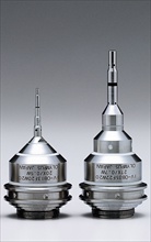 Olympus today released its unique MicroProbe Objective (MPO) lenses for studying the internal biology of living organisms. The two new water-immersion lenses, namely the 27x magnification IV-OB35F22W20 and the 20x IV-OB13F20W20, have an innovative needle-like design, with the lenses housed in tips measuring 3.5 mm and 1.3 mm in diameter. This means they can be inserted into very small surgical excisions, facilitating in vivo imaging without significantly disrupting the natural state of the tissue or organ being investigated...
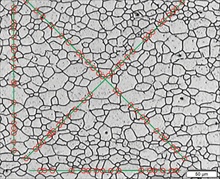 The OLYMPUS Stream Solutions enable you to easily capture and analyse any image for your niche applications quickly, precisely and in compliance with common international standards. Detailed in an educational demo video, the Grain Sizing Solutions are easy to use for beginners and experienced users alike, enabling you to accurately determine the quality of your steel. The Solutions are optional component to the OLYMPUS Stream software, which provides you with a completely intuitive workflow interface to accurately image and measure the microstructure of any sample....
Olympus releases the new CX22 series of microscopes for use in educational and routine microscopy
Jan 13, 2012
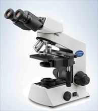 Olympus has today released the new CX22 microscopes for routine and educational microscopy. Building on the success of the CX21 series of microscopes, the CX22 series feature significant improvements for convenient microscopic observations. Without compromising on image quality, these microscopes provide a bigger field of view and increased flexibility when it comes to altering the height of the eye point. The quick and easy setting of different viewing heights makes this microscope series perfect for use in an educational setting where users are changing frequently...
 Olympus has released the new XLPLN25XSVMP SCALEVIEW 25x objective lens (NA 1.0) for the deep imaging of thick biological samples. The revolutionary SCALEVIEW approach was developed in collaboration with the RIKEN Brain Institute in Japan, where it allowed researchers to create highly accurate 3D structural representations of brain tissue. Until now, the use of such samples has been limited by the effects of tissue opacity and light scattering. To counter this, the new objective has a super-long-working-distance of 4mm and works alongside Olympus's new SCALEVEW-A2 clearing agent, which renders samples nearly translucent while preserving fluorescent signals...
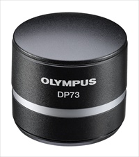 Olympus has today released the new ultra resolution DP73 and DP73WDR digital cameras, for quality without compromise in brightfield and fluorescence imaging. The new multi-purpose cameras leverage Olympus's expertise in pixel shift sensors and high-end consumer DSLR camera design to generate extremely detailed and accurately coloured images. This is achieved using the company's new ‘3CCD mode' and ‘Fine Detail Process' technologies. In addition, the DP73WDR employs the Olympus WiDER (Wide Dynamic Range) technology to ensure that each specific image area is always optimally exposed, regardless of any differences in intensity...
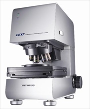 The Olympus LEXT OLS4000 is being used for the development of good practice guidelines and new calibration standards in optical metrology. These reference standards are being developed by a team led by Professor Richard Leach, Principal Research Scientist at the National Physical Laboratory (NPL). Preliminary data, presented by Professor Leach at the 10th International Symposium on Measurement Technology and Intelligent Instruments (ISMTII) demonstrated that these calibration standards will help users to...
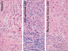 Olympus provides its versatile, powerful BX3 clinical microscope systems for analysis and disease diagnosis in pathology and cytology. The systems take advantage of Olympus's new true colour LED illumination technology and built in Light Intensity Manager (LIM) to create images with accurately rendered colours. Histological and cytological stains appear exactly the same under the true colour LED as they do under daylight filtered halogen...
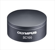 Olympus has introduced the new, easy-to-use SC100 digital colour camera for high quality brightfield imaging, especially where optimal colour reproduction and superior resolution are required. The 10.5 megapixels sensor of the SC100 allows samples to be investigated in minute detail, particularly when using a low magnification objective. This frees users from needing to take multiple, high magnification images of a sample to preserve resolution...
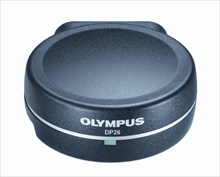 Olympus has released the new DP26, a five megapixel colour digital camera optimised for viewing, documentation, reporting and analysis using your microscope. The new camera is ideal for browsing a sample using the monitor, utilising a rapid progressive scan readout that provides fluid, natural panning and focusing, and avoids distracting artefacts. This removes the physical strain imposed by using the eyepieces, improving user comfort and efficiency...
 Olympus' new BX3 microscope systems are designed with the user in mind to maximise the quality of fluorescence images captured as part of life science research projects. The high-end BX63 system comfortably handles day-to-day applications, while boasting a range of advanced features to satisfy the most complex of needs, including rapid deconvolution of fluorescent Z-stack images...
 Olympus has released the latest version of its virtual slide (VS) product range, the VS120. The system can rapidly scan a slide in less than two minutes, creating a digital ‘virtual slide' that is an exact copy of the original. This is easily created and navigated using an intuitive graphical interface. The new unit provides semi-automatic and manual focusing modes for working with challenging samples, enhanced image clarity using the new Sample Detection Mask for automatic slide scanning, and increased optimisation of user workflow....
 Olympus has introduced two new silicon oil-immersion objectives, the UPLSAPO30xS and UPLSAPO60xS. The new lenses are ideal for live cell experiments, particularly those investigating thick samples or requiring long-term imaging. Users can generate bright images at higher resolution, as silicon oil significantly improves optical performance in comparison to conventional oil- or water-based approaches...
 Olympus attended the recent International Conference on Metrology and Properties of Engineering Surfaces, where it displayed its advanced measuring confocal laser scanning microscopy, the LEXT OLS4000. Hosted by the National Physical Laboratory (NPL), the event was highly successful with a record number of delegates in attendance...
 Olympus has introduced FV10-ASW 3.0, the latest version of its software for the FluoView FV1000 range of confocal laser scanning microscopes (cLSM) and Fluoview FV1000MPE (multiphoton excitation) systems. By incorporating high dynamic range imaging (HDRI), a greater dynamic range of fluorescence between the lightest and darkest areas of a sample is obtained. Furthermore, signal to noise ratios are minimised, enabling clear, highly resolved images to be easily captured...
 Olympus has today announced the release of the inverted IX81 microscope with ZDC2 Z-Drift compensation system. This next generation solution ensures that your samples are always in focus, providing extremely sharp images. The two modes, continuous and ‘one-shot', enable the completion of any time-lapse experiments...
 Olympus has launched the latest version of its highly successful Olympus Stream materials science microscopy imaging software family. Olympus Stream 1.6 provides a number of new and improved features, which not only increase workflow efficiency, but also enhance the overall capabilities of the software. Olympus Stream 1.6 is now compatible with the Microsoft Windows 7, 64 bit, operating system, making it fully compatible with all new computer systems...
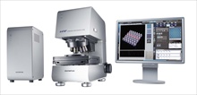 The Olympus LEXT OLS4000 is being used as a benchmark tool for the development of good practice guidelines and new reference standards in optical metrology. With both of these projects being detailed at the upcoming 13th International Conference on Metrology and Properties of Engineering Surfaces, hosted by the National Physics Laboratory (NPL) at Twickenham Stadium, Olympus is at the forefront of developments in metrology. The LEXT OLS4000 has therefore aided the development of various tools to improve the reliability and usability of optical metrology techniques...
The Olympus cellSens Dimension Count & Measure Solution module for automated image analysis
Mar 15, 2011
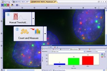 Olympus has announced the release of its Count & Measure Solution module for the cellSens Dimension software suite. The new module makes it simple to design and automate image analysis protocols by providing advanced object detection and structure differentiation. This maximises accuracy and reproducibility while minimising the time users must devote to manual image processing...
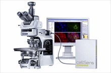 Olympus has announced the release of the BX63 microscope system, the top-of-the-range model of the powerful BX3 portfolio. BX3 microscope systems are built by your needs, with a modular, customisable design that makes them flexible enough to be adjusted to different experimental requirements, while providing inherent future-proof adaptability...
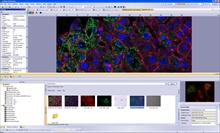 Olympus has today announced the release of its Database Solution module for the cellSens Dimension software suite. The module will simplify user workflow management, making it easier to capture, process and securely store images along with a variety of searchable and customisable metadata. With the ability to act as a client-server platform, indexed files and data can be shared across a network, with access and modification rights secured by password protection
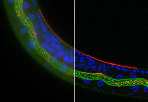 The Olympus cellSens deconvolution solution module employs a constrained iterative (CI) algorithm to remove out-of-focus blur in advanced life science imaging. This high-speed operation provides extremely sharp images, as well as near confocal quality, with the ability to correct for any spherical aberrations. Integrating seamlessly with the cellSens Dimension software, outstanding performance and quality are guaranteed
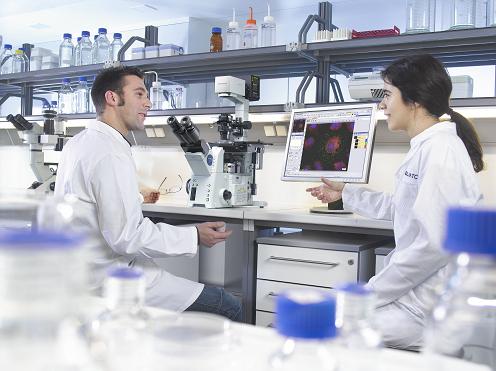 Olympus has now introduced labSens - a complete documentation package for clinical and research imaging. The software provides a workflow-oriented user interface, ensuring that the key functions for digital imaging, analysis and documentation are clearly presented and easy to use. This is achieved by seamlessly combining the new Olympus BX3 clinical microscopes with digital-imaging cameras, as well as automation components, to provide the perfect conduit between the user, the process and the microscope system.
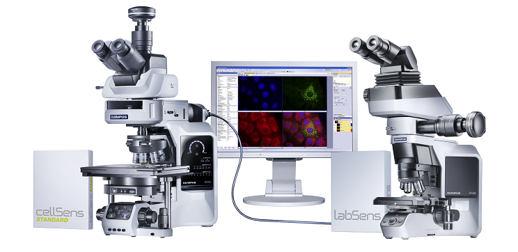 The new BX3 series sets a new standard in upright clinical and research microscopy. Thanks to its modularity the BX3 adapts to user requirements and applications even if they change over the years - an upright microscope system built by your needs.
Olympus releases v1.4 of its peerless Stream materials science microscopy imaging software
Sep 8, 2010
Olympus Introduces new DP21 digital microscopy camera for life-science and materials microscopy
Dec 11, 2009
Olympus enters cooperation to assist users of optical measurement equipment in deciding the best strategy
Dec 8, 2009
Media Partners