

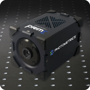
 Photometrics, a manufacturer of high performance scientific cameras for life science applications, today announces the release of its new sCMOS camera, the Photometrics Prime. Prime is the first 4.2 Megapixel scientific CMOS camera available for life science research that actively defeats the negative impact of Poisson noise in low light images.
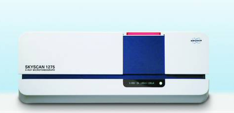 With the introduction of the new SKYSCAN™ 1275 highly automated, self-optimizing desktop X-ray microtomograph, Bruker puts class-leading imaging into the hands of materials and life scientists in both research and industrial applications, such as high-throughput 3D imaging in quality control and production process monitoring. The system takes advantage of new X-ray source technology and efficient flat-panel detectors to reduce...
 The minister was visiting the site at the Queen Elizabeth University Hospital where the new Imaging Centre of Excellence (ICE) will be based. The ICE, supported by £16m of UK Government funding through the Medical Research Council as part of the Glasgow & Clyde Valley City Deal, will provide clinical research facilities which will be unique in the UK and create more than 200 new jobs for local people. The centre is expected to be...
 Greiner Bio-One, a technology partner for the diagnostic and pharmaceutical industry, is unveiling its new CELLview slide at the European Biotechnica trade show in Hanover, Germany. The slide’s glass bottom is now available with both a TC-treated surface, for simple applications, and an Advanced TC-treated surface for more complex experiments such as multiple immunological staining or live cell analysis of sensitive cells...
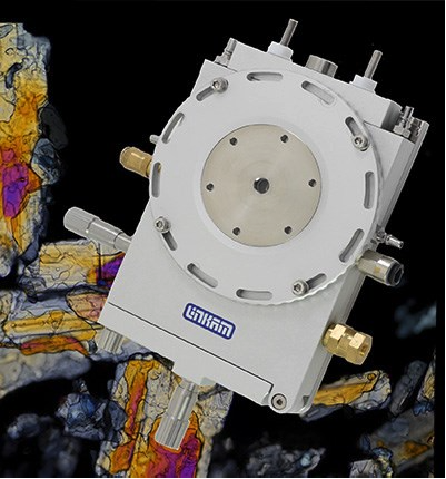 This work was carried out at the Katholic University of Leuven in Belgium. The Katholic University of Leuven in central Belgium provides nine clusters, or Key Areas, of scientific research. The Centre for Surface Chemistry and Catalysis is a group within the Department of Microbial and Molecular Systems, M2S. Fuel cells are one of the key areas of research. Fuel cells, and in particular polymer electrolyte membrane fuel cells (PEMFCs) have been earmarked as highly...
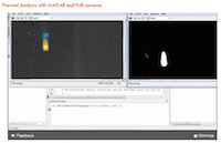 This new capability allows users to access their customized MatLab® scripts directly in ResearchIR for specially-tailored thermal image analysis and processing tasks. Many useful MatLab functions can now be directly accessed from within ResearchIR. Combining FLIR thermal and other cameras with MATLAB provides you with a flexible environment to explore algorithms for multi-sensor systems. To demonstrate the powerful utility of this new capability...
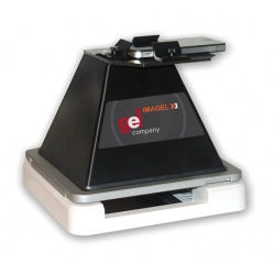 The Imagel can be used with 3 different light sources. Use the blue LED transilluminator for DNA applications that use Green DNA, GelGren, SYBR® Green, SYBR® Safe, SYBR® Gold and for Protein applications using LavaPurple
 Researchers in the Laboratory of Cell Culture at the Lithuanian University of Health Sciences Academy of Medicine are utilising the easy-to-use G:BOX Chemi XRQ system to image and analyse proteins on chemiluminescent Western blots and DNA gels stained with SYBR® Safe dyes. The image analysis data is being used to investigate the effects that genetic modification have on how stem cells differentiate into cardiac myocytes and integrate into cardiac...
 The company's comprehensive range of high resolution framing cameras, trajectory trackers, still cameras, image intensifiers and accessories open the door to exciting discoveries, ground breaking research and new measurement solutions in a wide range of applications areas. Visitors to the website are given open access to an informative bibliography of research articles, application notes, video clips and still images...
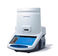 The Cell Counter model R1 is a cost-effective solution that saves time and simplifies the cell counting process. Engineered with leading-edge technology in a portable design, the new Olympus Cell Counter model R1 offers user-friendly and cost-effective cell counting for routine cell culturing. Accurate cell counts are important for a variety of research applications, including regenerative and drug medicine. Rather than relying on a human operator to manually count...
 This new capability allows users to access their customized MatLab® scripts directly in ResearchIR for specially-tailored thermal image analysis and processing tasks. Many useful MatLab functions can now be directly accessed from within ResearchIR. Combining FLIR thermal and other cameras with MATLAB provides you with a flexible environment to explore algorithms for multi-sensor systems. To demonstrate the powerful utility of this new capability...
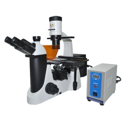 Eikonix, the Cambridge based imaging experts have recently introduced the MF50 Inverted Fluorescence Microscope to the UK market. This latest addition to the range of fluorescence microscopes and specialist blue/green LED lighting units gives customers an even greater choice when looking to purchase a high quality microscope unit at an affordable price.
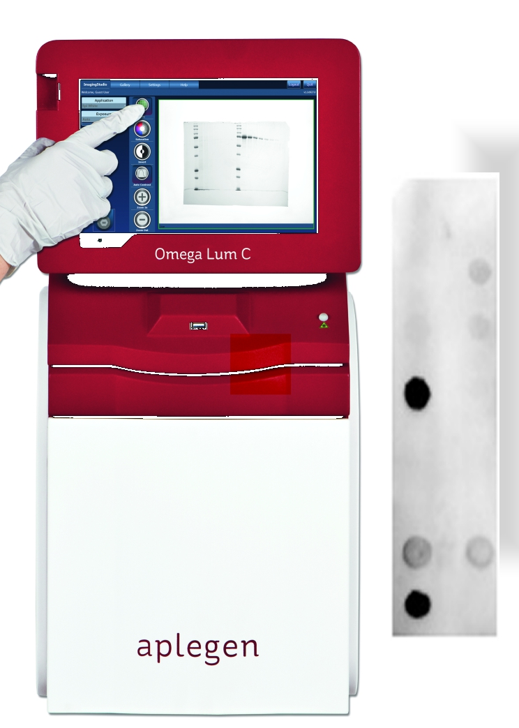 This chemiluminescent product joins the Company’s extensive range of over 800 reagents and consumables for the life scientist. Chemi FP has attomole sensitivity and a very long lasting signal output. The light emission is stable for 10 times longer than with typical ECL substrates. This now enables the user to detect bands not usually visualised with other substrates that are commonly used. Importantly, the high signal-to-noise level and large dynamic range of the product makes it ideal for quantifying low intensity bands.
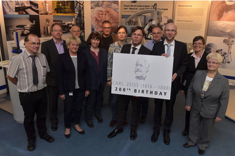 Carl Friedrich Zeiss was born in the German city of Weimar on 11 September 1816. The company ZEISS is celebrating the 200th birthday of its founding father by organizing many different activities and events – in addition to the Carl Zeiss Day on 11 September 2016 in downtown Jena, there will also be a touring exhibition and a book. ZEISS will celebrate its prominent founder together with players from the city of Jena, the ZEISS...
 The AutoTEC a120 is a second-generation, fully automated tissue embedder that eliminates the labor intensive need to manually orient and embed tissue specimens and form tissue or cell paraffin blocks. The AutoTEC technology, combined with the Paraform® Sectionable Cassette System ensure that the orientation of specimens is locked from grossing to microtomy for all routine tissue types, thereby eliminating the risk of orientation mistakes...
 Bruker today introduces four important new preclinical imaging systems at the World Molecular Imaging Congress 2015 in Honolulu, Hawaii. The novel products launched at WMIC each deliver improved performance and convenience for routine imaging, and open new horizons for advanced translational research, while Bruker’s platform philosophy facilitates multimodal imaging projects. Researchers will gain a more complete picture and...
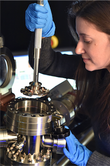 The US Naval Research Laboratory (NRL) has taken delivery of the US Department of Defense’s first Local Electrode Atom Probe (LEAP) microscope. The high- performance atom probe from CAMECA, a unit of AMETEK Materials Analysis, is used in advanced materials analysis to provide precise atom-by-atom identification, 3-D spatial positioning, and accurate atomic-scale reconstruction of a material’s microstructure. Since their development in the 1960s, atom probes have contributed to...
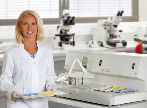 The independent Arcadia modules offer users the flexibility to organize the embedding workflow to their liking. HistoCore Arcadia is a combination of the paraffin dispensing module Arcadia H and the cold plate Arcadia C. Simple operation and precise control help improve the quality, reliability and speed of embedding work. The new station is designed with the user in mind and incorporates comfortable wrist-pads that increase comfort and stability...
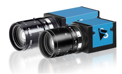 The Imaging Source, an international manufacturer of machine vision cameras, has just announced the immediate availability of new industrial cameras with the Sony Full HD WDR Sensor IMX236. The cameras feature compact, robust industrial housing with C/CS- or S-Mount and are available as GigE (PoE) and USB 3 versions in monochrome and color. With integrated WDR (Wide Dynamic Range) and a resolution from VGA to Full HD, they are...
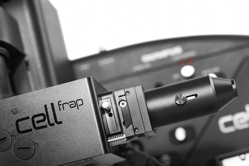 Enabling the easy addition of photomanipulation techniques to imaging platforms, Olympus has launched a new cellFRAP deck system for the popular IX3 ‘open source concept’ IX83 and IX73 microscopes. Olympus cellFRAP is designed with researchers in mind, providing highly accurate and flexible live-cell photomanipulation with various evaluation options for data presentation needs. Unlike conventional widefield-based FRAP systems, Olympus’ cellFRAP...
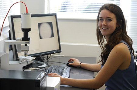 Linkam's continued growth in sales worldwide has led to a further hire in the support team under sales and marketing manager, Duncan Stacey. He outlines the position: “With our sales and product development being driven by feedback from our users a Market leaders in temperature controlled microscopy, Linkam Scientific Instruments, are pleased to announce a new member of staff to join their growing sales and marketing support team. Linkam's continued...
 Nanolab Technologies Inc., a Silicon Valley-based analytical services lab, has purchased a new Local Electrode Atom Probe from CAMECA Instruments Inc. The high-performance atom probe from CAMECA, a unit of the AMETEK Materials Analysis Division, is used to provide advanced materials analysis, including precise atom-by-atom identification, 3-D spatial positioning, and accurate atomic-scale reconstruction of a material’s...