Researchers have developed a family of engineered nanobodies which neutralise the SARS-CoV-2 virus, targeting the viral spike protein in a novel way. The research team from The Rosalind Franklin Institute, working with colleagues at Diamond Light Source, Oxford University and Public Health England, have hailed the breakthrough as a potential therapy....
The ZEISS Innovation Hub on the campus of the Karlsruhe Institute of Technology (KIT) has seen a number of successful collaborations and projects since it opened in early 2020. ZEISS wants the hub to house high-tech and digital start-ups, as well as its own innovation and new business activities. KIT will thus join forces with ZEISS experts to pave the way for the technologies of the future....
The launch of a new JEOL Field Emission Scanning Electron Microscope during the summer of 2020 includes virtual demonstrations of its powerful performance directly to those in the market for an analytical ultrahigh resolution SEM. JEOL’s new JSM-IT800 is the company’s top-of-the-line microscope with ultrahigh spatial resolution imaging and analysis at the nanoscale....
BioTek Instruments announces the availability of variable bandwidth monochromators on their modular and versatile Synergy™ H1 Hybrid Multi-Mode Reader. This allows researchers to achieve even greater levels of assay sensitivity and specificity compared to fixed bandwidth systems. The microplate reader includes BioTek’s patented Hybrid Technology™...
A radical new way of thinking about soil has finally solved the mystery of why adding organic material like manure improves flood and drought resilience, climate control and crop yields - universal ‘ecosystem services’ that are widely recognised as worth billions to the global economy. Founded on more than 50 years’ worth of data from a unique field experiment, researchers have demonstrated...
The new SCHOTT® VisiLED UV Ring Light for stereo microscopes combines classic bright-field illumination with UV illumination. It is the only segment ring light on the market in which white-light and UV-LEDs are alternately installed in eight segments. This LED arrangement allows objects to be examined from the same illumination angle, which significantly improves the ability to compare and reproduce resulting images....
NanoImaging Services, Inc. (NIS), specialists in transmission electron microscopy (TEM) with a vision to make cryoEM workflows accessible to all, has announced the opening of a new facility in Boston. NIS is expanding with new services and locations to support its growing customer base requiring cryoEM for drug discovery and vaccine development applications. COVID-19 projects are receiving prioritization....
3D label-free imaging identifies real-time cholesterol sorting mechanism in Plasmodium falciparum-infected human erythrocytes. The first study to elucidate the sequential dynamics of membrane cholesterol transport in erythrocytes infected with live Plasmodium falciparum parasites has been successfully concluded using the 3D label-free imaging capability of holotomography microscopy....
Rutherford has entered into a £55m development framework agreement with Equitix Limited. Under of the terms of the agreement, Rutherford and Equitix will establish up to five new diagnostic facilities in the UK, to provide diagnostic services to the NHS and to private patients. Each centre will provide a variety of diagnostics services including Positron Emission Tomography–Computed Tomography, Magnetic Resonance Imaging, Computed Tomography, Ultrasound, Endoscopy, X-Ray and other relevant diagnostic services...
CytoSMART Technologies has announced the launch of a new live-cell imaging system. The CytoSMART Lux2 Duo Kit offers a straightforward, cost-effective solution for researchers carrying out immediate side-by-side comparisons between cell cultures. Said Jan-Willem van Bree, CTO at CytoSMART Technologies "Employing this two camera mini live cell imaging system is especially useful for stem cell research....
Having launched a unique AI-aided drug discovery platform last month, this company grows quickly with concerted support from many talented researchers and scientists. It offers drug R & D solutions from the perspective of AI for medical institutions and pharmaceutical enterprises worldwide...
Oxford Instruments and Digital Surf, creator of the industry-standard Mountains® surface and image analysis software platform, has announced the release of Relate software for users of Oxford Instruments' leading-edge tools for materials characterization. This software will bring great value to Oxford Instruments' users working in R&D across a wide range of academic and industrial applications including semiconductors, renewable energy, mining, metallurgy, and forensics....
In this new 10-page Technology Note, learn how different image processing methods have the potential to make widefield microscope systems more powerful and versatile. Understand each methods limitations and pitfalls, as well as suitability for your specific applications. For decades, fluorescence microscopy has been an invaluable tool in life sciences research, with new variations and implementations emerging almost every year....
Indica Labs, a leading provider of computational pathology software, and Octo, a premiere information technology systems provider to the U.S. Federal Government, are pleased to announce the online COVID Digital Pathology Repository (COVID-DPR), a virtual collection of high resolution microscopic COVID-related human tissue images hosted at the National Institutes of Health....
When UCLA’s Dr. Araceli Espinosa-Jeffrey sent human brain cells into space, her goal was to gain a better understanding of how neural stem cells grow and develop in microgravity. That understanding is key to discovering more about the serious issues of intracranial hypertension affecting astronauts returning from space. The information may also one day be used to further cell replacement therapies...
Jian-Wei Pan, Professor at the University of Science and Technology of China, in Hefei, is the winner of the prestigious ZEISS Research Award 2020. He is one of the world's leading researchers in the field of quantum technology. One of the most remarkable results of Jian-Wei Pan's research is the distribution of entangled photons over a distance of 1,200 km, by far the longest distance ever reached....
To capture the importance of this initiative, and in anticipation of the grand opening of the Ellison Institute’s new building, Olympus has created a video, highlighting the relationship between imaging analysis tools and precision cancer medicine. One focus of the partnership is encouraging projects that engage translational oncology and precision anti-cancer drug screening...
Linkam has recently seen an increased interest in its stage that was specifically designed for the analysis of catalytic reactions: the CCR1000. Catalysis is a technique for improving the yield or rate of a chemical reaction using a catalyst material. A wide range of catalytic materials are available; including aluminosilicates, which are used in the petrochemical industry to reduce natural materials to smaller hydrocarbons...
The sensitivity of Beckman Coulter Life Sciences’ CytoFLEX Flow Cytometer is underpinning a pioneering multinational research initiative. This is designed to transform the lives of those suffering from a disfiguring and potentially life threatening parasitic disease. The research team has just secured a grant of eight million Euros from the European & Developing Countries Clinical Trials Partnership...
Vision Engineering, a 61 year old British leading designer and manufacturer of high-quality visual measurement and inspection technologies, has attained ISO 17025:2017 accreditation from UKAS and is now a UKAS accredited calibration laboratory No. 7706. The award of ISO 17025:2017 by UKAS Accreditation, which is globally recognised, is the only mechanism that determines the technical competence and integrity of the organisations offering testing and calibration services....
For use in advanced medical research into inflammation, oncology and nephritis the 7T PET-MR imaging system from MR Solutions provides a combination of PET and MRI images either simultaneously or separately. This ground breaking combination technology allows researchers to have superior soft tissue contrast and molecular imaging together.
Olympus cellSens imaging software improves the efficiency of your research with accurate object detection and segmentation. Leveraging the power of deep learning, Olympus cellSens imaging software for microscopy offers significantly improved segmentation analysis, such as label-free nucleus detection and cell counting, for more accurate data and efficient experiments....

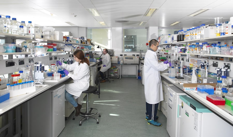 Researchers have developed a family of engineered nanobodies which neutralise the SARS-CoV-2 virus, targeting the viral spike protein in a novel way. The research team from The Rosalind Franklin Institute, working with colleagues at Diamond Light Source, Oxford University and Public Health England, have hailed the breakthrough as a potential therapy....
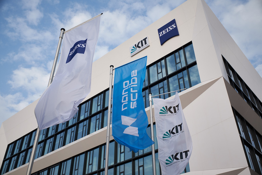 The ZEISS Innovation Hub on the campus of the Karlsruhe Institute of Technology (KIT) has seen a number of successful collaborations and projects since it opened in early 2020. ZEISS wants the hub to house high-tech and digital start-ups, as well as its own innovation and new business activities. KIT will thus join forces with ZEISS experts to pave the way for the technologies of the future....
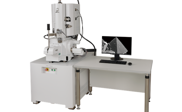 The launch of a new JEOL Field Emission Scanning Electron Microscope during the summer of 2020 includes virtual demonstrations of its powerful performance directly to those in the market for an analytical ultrahigh resolution SEM. JEOL’s new JSM-IT800 is the company’s top-of-the-line microscope with ultrahigh spatial resolution imaging and analysis at the nanoscale....
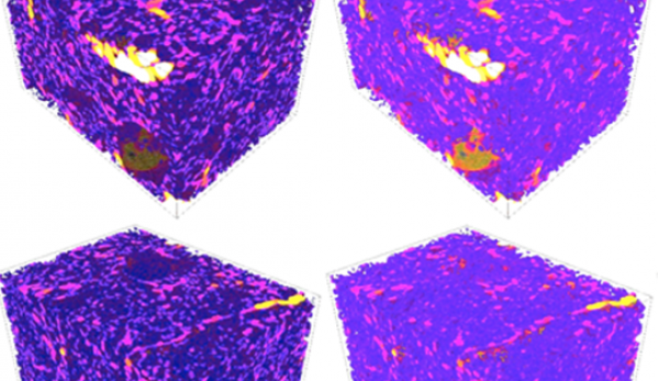 A radical new way of thinking about soil has finally solved the mystery of why adding organic material like manure improves flood and drought resilience, climate control and crop yields - universal ‘ecosystem services’ that are widely recognised as worth billions to the global economy. Founded on more than 50 years’ worth of data from a unique field experiment, researchers have demonstrated...
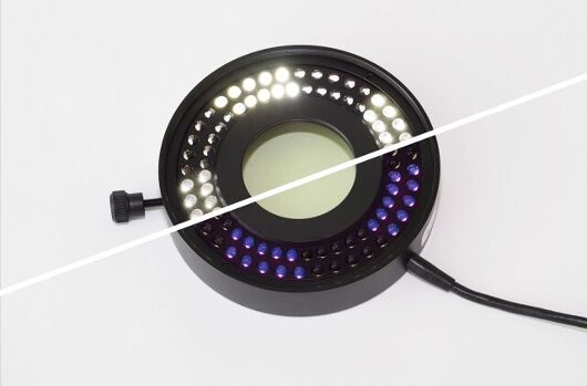 The new SCHOTT® VisiLED UV Ring Light for stereo microscopes combines classic bright-field illumination with UV illumination. It is the only segment ring light on the market in which white-light and UV-LEDs are alternately installed in eight segments. This LED arrangement allows objects to be examined from the same illumination angle, which significantly improves the ability to compare and reproduce resulting images....
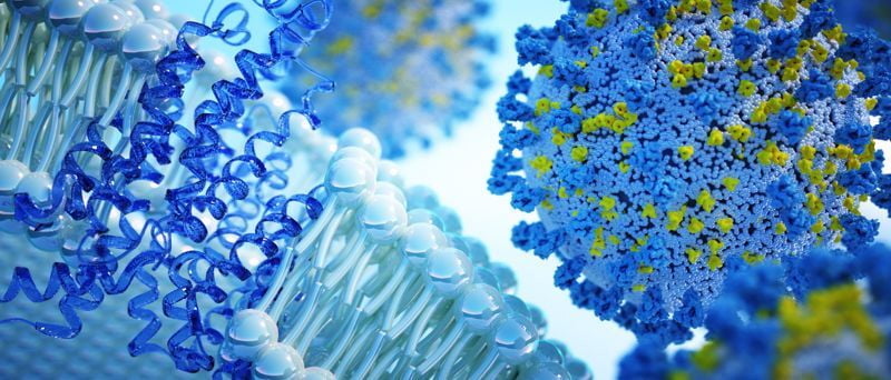 NanoImaging Services, Inc. (NIS), specialists in transmission electron microscopy (TEM) with a vision to make cryoEM workflows accessible to all, has announced the opening of a new facility in Boston. NIS is expanding with new services and locations to support its growing customer base requiring cryoEM for drug discovery and vaccine development applications. COVID-19 projects are receiving prioritization....
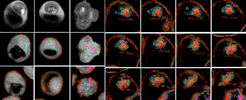 3D label-free imaging identifies real-time cholesterol sorting mechanism in Plasmodium falciparum-infected human erythrocytes. The first study to elucidate the sequential dynamics of membrane cholesterol transport in erythrocytes infected with live Plasmodium falciparum parasites has been successfully concluded using the 3D label-free imaging capability of holotomography microscopy....
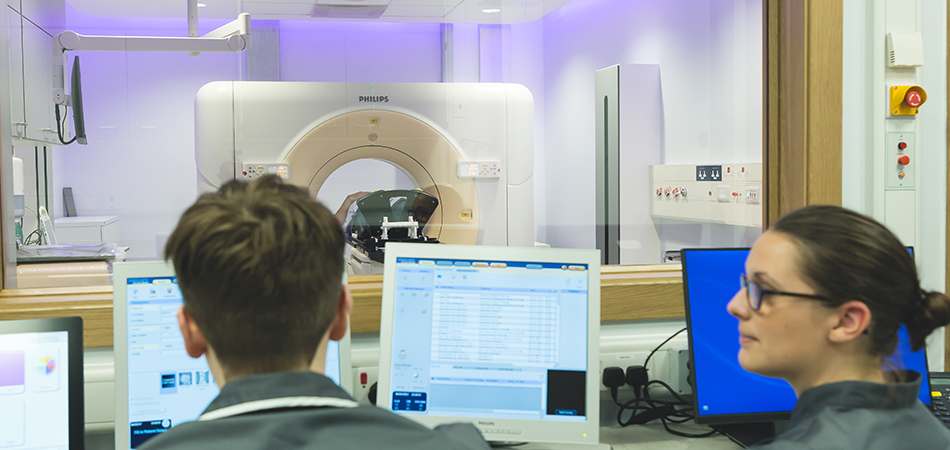 Rutherford has entered into a £55m development framework agreement with Equitix Limited. Under of the terms of the agreement, Rutherford and Equitix will establish up to five new diagnostic facilities in the UK, to provide diagnostic services to the NHS and to private patients. Each centre will provide a variety of diagnostics services including Positron Emission Tomography–Computed Tomography, Magnetic Resonance Imaging, Computed Tomography, Ultrasound, Endoscopy, X-Ray and other relevant diagnostic services...
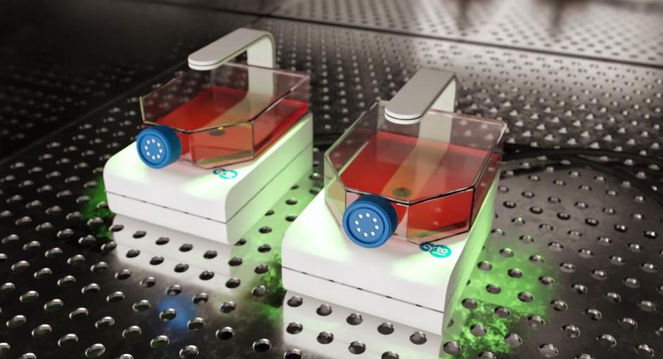 CytoSMART Technologies has announced the launch of a new live-cell imaging system. The CytoSMART Lux2 Duo Kit offers a straightforward, cost-effective solution for researchers carrying out immediate side-by-side comparisons between cell cultures. Said Jan-Willem van Bree, CTO at CytoSMART Technologies "Employing this two camera mini live cell imaging system is especially useful for stem cell research....
 Having launched a unique AI-aided drug discovery platform last month, this company grows quickly with concerted support from many talented researchers and scientists. It offers drug R & D solutions from the perspective of AI for medical institutions and pharmaceutical enterprises worldwide...
 Oxford Instruments and Digital Surf, creator of the industry-standard Mountains® surface and image analysis software platform, has announced the release of Relate software for users of Oxford Instruments' leading-edge tools for materials characterization. This software will bring great value to Oxford Instruments' users working in R&D across a wide range of academic and industrial applications including semiconductors, renewable energy, mining, metallurgy, and forensics....
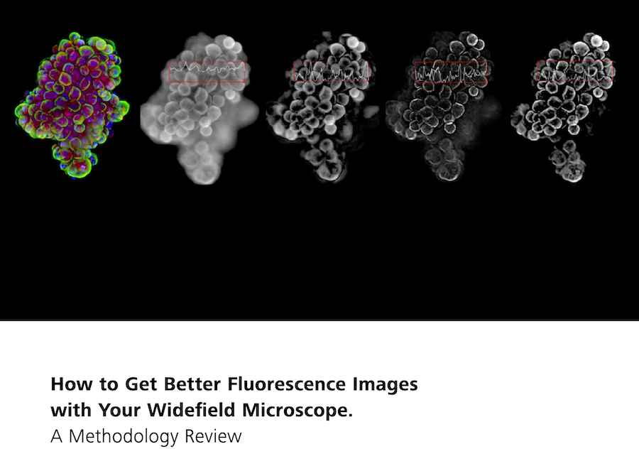 In this new 10-page Technology Note, learn how different image processing methods have the potential to make widefield microscope systems more powerful and versatile. Understand each methods limitations and pitfalls, as well as suitability for your specific applications. For decades, fluorescence microscopy has been an invaluable tool in life sciences research, with new variations and implementations emerging almost every year....
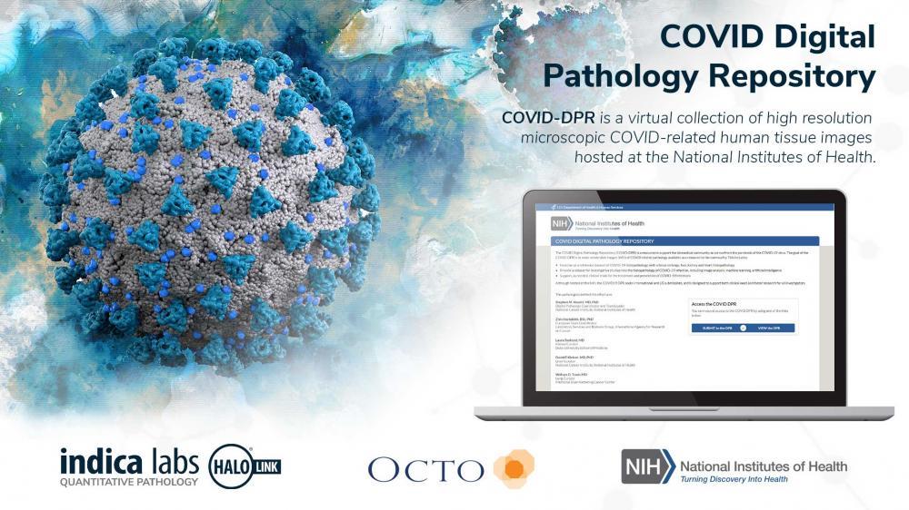 Indica Labs, a leading provider of computational pathology software, and Octo, a premiere information technology systems provider to the U.S. Federal Government, are pleased to announce the online COVID Digital Pathology Repository (COVID-DPR), a virtual collection of high resolution microscopic COVID-related human tissue images hosted at the National Institutes of Health....
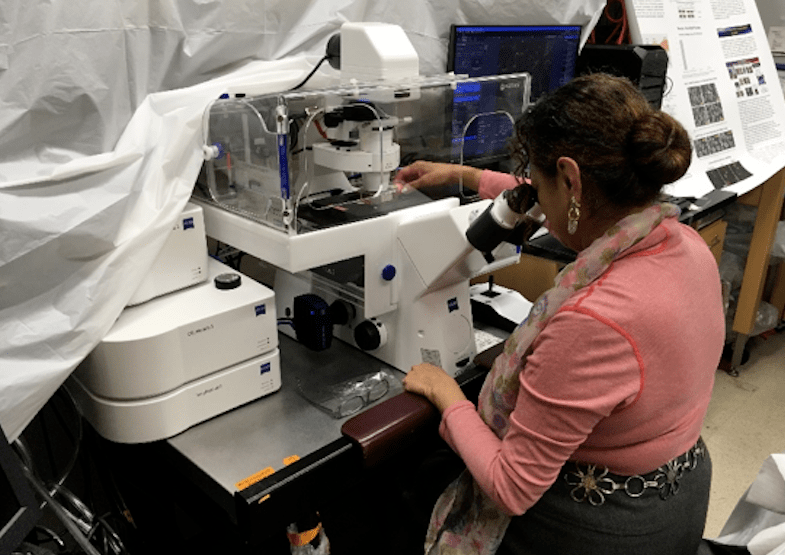 When UCLA’s Dr. Araceli Espinosa-Jeffrey sent human brain cells into space, her goal was to gain a better understanding of how neural stem cells grow and develop in microgravity. That understanding is key to discovering more about the serious issues of intracranial hypertension affecting astronauts returning from space. The information may also one day be used to further cell replacement therapies...
 Jian-Wei Pan, Professor at the University of Science and Technology of China, in Hefei, is the winner of the prestigious ZEISS Research Award 2020. He is one of the world's leading researchers in the field of quantum technology. One of the most remarkable results of Jian-Wei Pan's research is the distribution of entangled photons over a distance of 1,200 km, by far the longest distance ever reached....
 To capture the importance of this initiative, and in anticipation of the grand opening of the Ellison Institute’s new building, Olympus has created a video, highlighting the relationship between imaging analysis tools and precision cancer medicine. One focus of the partnership is encouraging projects that engage translational oncology and precision anti-cancer drug screening...
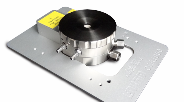 Linkam has recently seen an increased interest in its stage that was specifically designed for the analysis of catalytic reactions: the CCR1000. Catalysis is a technique for improving the yield or rate of a chemical reaction using a catalyst material. A wide range of catalytic materials are available; including aluminosilicates, which are used in the petrochemical industry to reduce natural materials to smaller hydrocarbons...
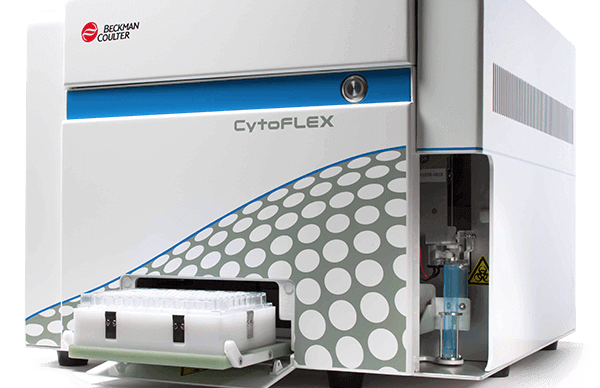 The sensitivity of Beckman Coulter Life Sciences’ CytoFLEX Flow Cytometer is underpinning a pioneering multinational research initiative. This is designed to transform the lives of those suffering from a disfiguring and potentially life threatening parasitic disease. The research team has just secured a grant of eight million Euros from the European & Developing Countries Clinical Trials Partnership...
 Vision Engineering, a 61 year old British leading designer and manufacturer of high-quality visual measurement and inspection technologies, has attained ISO 17025:2017 accreditation from UKAS and is now a UKAS accredited calibration laboratory No. 7706. The award of ISO 17025:2017 by UKAS Accreditation, which is globally recognised, is the only mechanism that determines the technical competence and integrity of the organisations offering testing and calibration services....
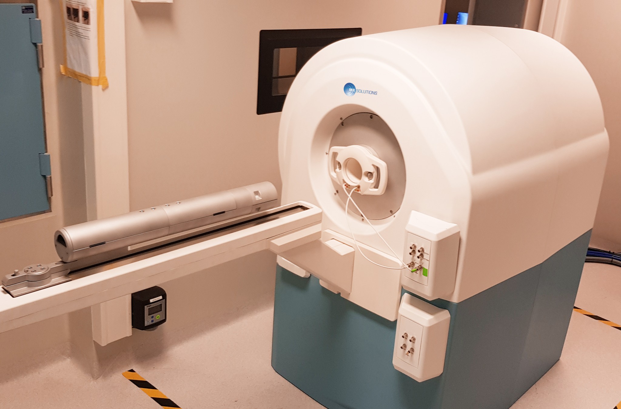 For use in advanced medical research into inflammation, oncology and nephritis the 7T PET-MR imaging system from MR Solutions provides a combination of PET and MRI images either simultaneously or separately. This ground breaking combination technology allows researchers to have superior soft tissue contrast and molecular imaging together.
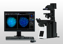 Olympus cellSens imaging software improves the efficiency of your research with accurate object detection and segmentation. Leveraging the power of deep learning, Olympus cellSens imaging software for microscopy offers significantly improved segmentation analysis, such as label-free nucleus detection and cell counting, for more accurate data and efficient experiments....