ZEISS and BOSELLO HIGH TECHNOLOGY have announced that the ZEISS Group will acquire a majority stake in the provider of industrial X-ray solutions.
For ZEISS, BOSELLO’s tailor-made solutions are a further step in the process of evolving into a one-stop provider of non-destructive measuring and inspection technology. The shared goal is to strengthen inline computed tomography....
FLIR Systems A6260sc camera sets the standard for Short Wavelength Infrared (SWIR) cameras for science and R&D applications by combining high-speed performance with fully customizable features.
Incorporating a proprietary high-resolution detector, the A6250sc camera offers improved sensitivity and linearity across the full dynamic range, making it ideal for radiometry and temperature calibrated applications....
Syngene is delighted to announce its GeneSys software has been upgraded to include QuickQuant, a new feature for rapid band quantification analysis on blots or gels.
This software, available to download free of charge to existing users of specified Syngene imaging systems, saves scientists time by allowing them to accurately analyse gels and blots at the point of image capture. For use in Syngene’s G:BOX and GeneGnome systems, QuickQuant is ideal for scientists who need to perform rapid band quantification while they are using their imager....
FLIR Systems A6260sc camera sets the standard for Short Wavelength Infrared (SWIR) cameras for science and R&D applications by combining high-speed performance with fully customizable features.
Incorporating a proprietary high-resolution detector, the A6250sc camera offers improved sensitivity and linearity across the full dynamic range, making it ideal for radiometry and temperature calibrated applications. The A6260sc is equipped with an indium gallium arsenide (InGaAs) detector....
Syngene is delighted to introduce its new GeneGnome XRQ, a dedicated chemiluminescence imaging system designed to rapidly and accurately image chemiluminescent Western blots.
The new, blue GeneGnome XRQ houses a high-quality F/0.95 fixed focus, cooled camera with on chip integration and GeneSys software, all packed into a light-tight darkroom. Based on the optimised short ‘camera to sample’ distance technology of the award winning GeneGnome....
The partnership makes Cytosurge's FluidFM technology available on the JPK NanoWizard AFM platform.
JPK Instruments, a world-leading manufacturer of nanoanalytic instrumentation for research in life sciences and soft matter, announce an exciting partnership with the Swiss company Cytosurge AG....
Horizon 2020 awarded to expedite the development of a 1GPixel resolution ultra-large format aerial surveillance camera.
Horizon 2020 is the biggest EU Research and Innovation programme ever with nearly €80 billion of funding available over 7 years (2014 to 2020). Horizon 2020 is a flagship initiative aimed at securing Europe's global competitiveness....
Introducing CorrStub™ - a unique range of SEM specimen stubs designed specifically for repeatable analysis over the same reference point
Combinations of techniques such as SEM and light microscopy are often used to determine complete characterisation of specimens. With this in mind Agar Scientific have developed the new CorrStub™ specimen stub....
Prior Scientific is pleased to announce the launch of its newly designed website.
The new website features user friendly navigation, improved functionality, enhanced rich content and a clean uncluttered design. The website will be consistently updated with product launches, business activity, corporate milestones and events....
Saving time with a four times higher scan speed, Olympus’ new confocal microscope improves efficiency in industrial inspection.
The LEXT OLS5000 combines 4K scanning and a large working distance with intuitive software to capture the shape of any sample under any angle. Olympus’ LEXT OLS5000 3D measuring laser microscope helps users reach the next level of metrology....
Phasefocus continues to increase its global international profile with the appointment of Nuohai Life Sciences as the company’s exclusive distributor for mainland China, Hong Kong and Macau.
Nuohai Life Sciences will provide sales, service and technical support for the Livecyte™ Cell Imaging System throughout the region, building on their reputation for introducing innovative new technologies for the Life Science Industry into the Chinese market....
First ZEISS ZEN Intellesis solution enables segmentation of correlative microscopy datasets
ZEISS announces ZEISS ZEN Intellesis, a new machine learning capability that enables researchers to perform advanced analysis of their imaging samples across multiple microscopy methods. The first algorithmic solution introduced by the ZEISS ZEN Intellesis platform makes...
NanoHybrids has been awarded $300,000 by the National Cancer Institute of the National Institutes of Health (NIH) to enhance the therapeutic efficacy of photodynamic therapy (PDT) in oral cancers using a novel nano-enabled hybrid technology.
The joint project will involve important in vivo pre-clinical studies designed to validate the efficacy of a nano-enabled platform to raise the therapeutic index of PDT in oral cancer. The nano-enabled hybrid technology’s relevance derives from an ability to enhance potency.... Read MoreNew Field Emission Scanning Electron Microscope ZEISS GeminiSEM 450 IntroducedDec 22, 2017
Addressing the highest demands in imaging and analytics from any sample, ZEISS introduces its new field emission scanning electron microscope (FE-SEM) ZEISS GeminiSEM 450. The instrument combines ultrahigh resolution imaging with the capability to perform advanced analytics while maintaining flexibility and ease-of-use.
Sectra announces that all St. Antonius Hospital locations have now gone live with Sectra’s enterprise image management solution for radiology, nuclear medicine and cardiology. With this solution for handling medical images across multiple locations and medical disciplines in a single system, physicians at St. Antonius can seamlessly share images and information with each other. Using a single system that is tightly integrated with the Epic EMR system will also reduce the workload of the IT department....
Grundium is developing the first portable digital microscope scanner in the world
China based investment firm, Ascend Capital Partners, has invested two million Euros to help Grundium OY develop their innovative portable digital microscope scanner. Grundium’s vision is to bring digital microscope scanners to every pathologist’s desk.....
APAS® is a breakthrough artificial intelligence technology for the automated imaging, image analysis, interpretation and reporting of growth on microbiology plates after incubation. The APAS® Independence, which is about the size of a large photocopier, improves the diagnostic efficiency of microbiology laboratories and enables faster reporting of infectious diseases
Deben reports on a publication in Microscopy & Microanalysis from the La Trobe Institute for Molecular Science.
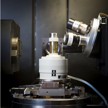
This shows a Deben CT500 tensile and compression stage being used in conjunction with a ZEISS Xradia Micro XCT system to enable a better understanding of ceramic matrix composites under various tensile bending loads. Dr Benedicta Arhatari is X-ray Tomography Laboratory Manager....
JPK Instruments reports on the exciting research of the Kim Group in the Structural Biology Laboratory of the Sungkyunkwan University School of Medicine, (SKKU), Suwon, South Korea using JPK's NanoWizard® ULTRA Speed AFM to study the binding of transcription factor Sox2 with super enhancers.
Professor Kyeong Kyu Kim leads a research group in the Structural Biology Laboratory of Sungkyunkwan University (SKKU) in Suwon, South Korea. The goal of their research is to understand the working mechanisms of Sox2, a master transcription factor that plays a role in controlling the “stemness” of cells....
Specialised Imaging has developed a new colour version of its SIR3 camera.
Capable of capturing up to11-million-pixel (4008 x 2688) resolution images in 36-bit colour across a wide dynamic range, the SIR3 colour camera enables amazing colorimetric effects on fast moving objects to be recorded clearly for the first time. Employing red, green and blue filters on each image intensifier channel....
High resolution 3D block face imaging for biological samples with fast acquisition rates and minimal sample damage
In collaboration with the National Center for Microscopy and Imaging Research (NCMIR) at the University of California San Diego, ZEISS releases a new Focal Charge Compensation module for block face imaging with ZEISS GeminiSEM and 3View® from Gatan, Inc....
Improved optical sectioning delivers higher resolution without the need to acquire a z-stack
At Neuroscience 2017, a new imaging mode for the ZEISS LSM 8 family with Airyscan has been introduced. Their unique 32-channel GaAsP array detector captures more spatial information than traditional confocal microscopes....


 ZEISS and BOSELLO HIGH TECHNOLOGY have announced that the ZEISS Group will acquire a majority stake in the provider of industrial X-ray solutions.
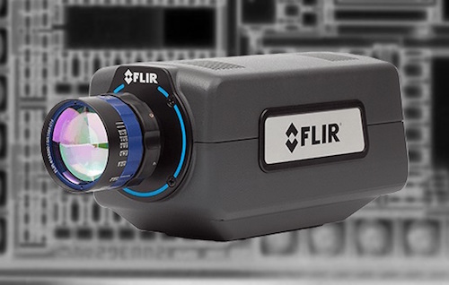 FLIR Systems A6260sc camera sets the standard for Short Wavelength Infrared (SWIR) cameras for science and R&D applications by combining high-speed performance with fully customizable features.
 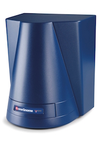 Syngene is delighted to introduce its new GeneGnome XRQ, a dedicated chemiluminescence imaging system designed to rapidly and accurately image chemiluminescent Western blots.
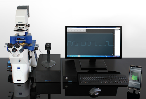 The partnership makes Cytosurge's FluidFM technology available on the JPK NanoWizard AFM platform.
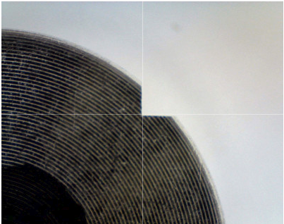 Introducing CorrStub™ - a unique range of SEM specimen stubs designed specifically for repeatable analysis over the same reference point
 Prior Scientific is pleased to announce the launch of its newly designed website.
 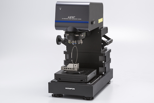 Saving time with a four times higher scan speed, Olympus’ new confocal microscope improves efficiency in industrial inspection.
  Phasefocus continues to increase its global international profile with the appointment of Nuohai Life Sciences as the company’s exclusive distributor for mainland China, Hong Kong and Macau.
 First ZEISS ZEN Intellesis solution enables segmentation of correlative microscopy datasets
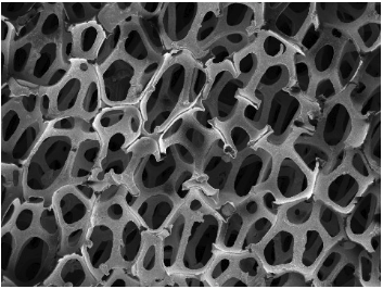

 Deben reports on a publication in Microscopy & Microanalysis from the La Trobe Institute for Molecular Science.
 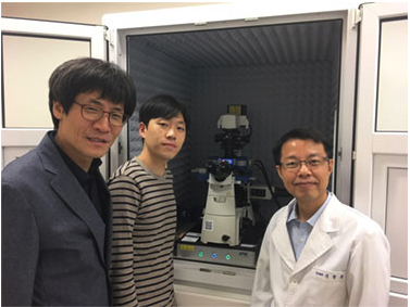 JPK Instruments reports on the exciting research of the Kim Group in the Structural Biology Laboratory of the Sungkyunkwan University School of Medicine, (SKKU), Suwon, South Korea using JPK's NanoWizard® ULTRA Speed AFM to study the binding of transcription factor Sox2 with super enhancers.
 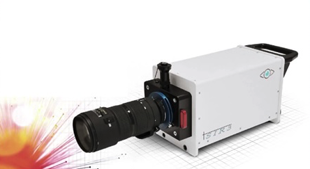 Specialised Imaging has developed a new colour version of its SIR3 camera.
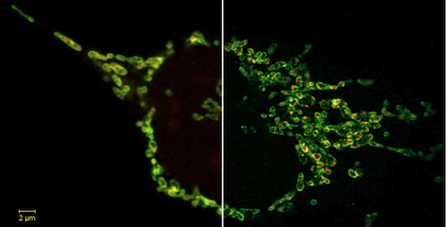 Improved optical sectioning delivers higher resolution without the need to acquire a z-stack