Next Generation Atom Probe Included Among 100 Most-Significant Developments in Research and Development
LEAP 5000 Atom Probe Offers Precise Atom-by-Atom, Sub-Nanometric Analysis of Metals, Semiconductors and Other Materials
CAMECA, a unit of AMETEK Materials Analysis, was recognized among the winners of the 54th Annual R&D 100 Awards by the editors of R&D magazine for its development of the LEAP 5000. Launched in August 2014, the LEAP 5000 is the latest generation of atom probe microscopes, which offer precise atom-by-atom identification, 3-D spatial positioning, and accurate atomic-scale reconstruction of a material’s microstructure...
Spectronics Corporation produces state-of-the-art products that technicians need to perform everyday essential laboratory applications.
One such product is Spectroline®CX-20 high-intensity UV viewing cabinet. Designed for peak efficiency, it guarantees maximum ultraviolet irradiance and fluorescent contrast. The CX-20 cabinet combines separate long-wave and short-wave 8-watt UV light sources with uniquely designed specular aluminum reflectors to assure maximum intensity and exceptional fluorescent contrast. An internal 25-watt white light bulb provides visible illumination...
EM Resolutions, manufacturers and suppliers of tools and accessories for users of electron microscopes, announce the availability of EM-Tec FIB grids which offer a secure way to attach TEM lamellas to the posts during lift-out procedures with FIB or SEM/FIB systems.
The new EM-Tec range of FIB lift-out grids from EM Resolutions provides a secure way to attach TEM lamella prepared with Focussed Ion Beam (FIB) instruments. Available in multiple post configurations, with a shape optimized for easy accessibility, EM-Tec FIB grids are compatible with all standard 3 mm TEM grid holders. EM-Tec FIB lift-out grids are available in three types: copper, molybdenum and unique, smooth-walled molybdenum...
Delivering new benchmarks in dynamic performance and accuracy, Queensgate’s NPC-D-5200 is a digital controller for use with the company’s highly advanced closed loop piezo actuators and stages.
Queensgate pioneered the use of capacitive sensors to provide precise positional feedback in closed loop; the NPC-D-5200 incorporates a precision capacitive measurement circuit and is updated with the stage position 120000 times per second, delivering high positional accuracy at speed. It has the capacity to address a broad spectrum of highly demanding alignment and metrology applications. By employing proprietary low noise...
The funds will be used to fuel international growth and to enter into the in-vitro diagnostics market.
Ovizio Imaging Systems, an innovative Belgian company developing cell counting solutions based on quantitative microscopy for life sciences applications, today announces it has secured a funding round of €8m ($9.1m) co-led by New Science Ventures, a US-based venture capital firm, and a private investor. This funding round...
LaVison BioTec, developers of advanced microscopy solutions for the life sciences, report on the work of Nicolas Renier, a Post-Doctoral Fellow in the laboratory of Marc Tessier-Lavigne at the Rockefeller University in New York where he applies light sheet microscopy to study axon rewiring in adult nervous systems.
Drs Nicolas Renier and Zhuhao Wu are post-doctoral fellows in the laboratory of Marc Tessier-Lavigne at the Rockefeller University in New York where they have co-developed methodologies to develop new imaging techniques applying light sheet microscopy to the study axon rewiring in adult nervous systems. It is hoped that this could lead to an understanding as to how experience or even pathologies such as Alzheimer's...
Complete workflow automation for long term cell assays
BioTek Instruments released the BioSpa™ 8 Automated Incubator, a unique platform that links microplate washers and dispensers with readers and imaging systems for unattended workflow automation. Real time temperature and CO2/O2 control and monitoring, plus humidity level monitoring and plate lid handling provide an ideal environment for cell-based and other assays, with minimal manual intervention...
Market leaders in temperature controlled microscopy, Linkam Scientific Instruments report on the use of their temperature controlled stages applied to CLEM and fluorescence microscopy to assist in endocytic sorting in the School of Biochemistry at the University of Bristol.
Dr Paul Verkade is a Reader in Cell Imaging in the School of Biochemistry at the University of Bristol where he also heads the Electron Microscopy unit of the Wolfson Bioimaging Facility. His current research focus is to develop techniques and tools for the use of Correlative Light Electron Microscopy (CLEM) studying endocytic sorting. Dr. Verkade is currently chair of the Electron Microscopy section of the Royal Microscopy Society and chair of...
Mazurek Optical Services are pleased to announce they have recently installed a suite of light microscopes in the School of Earth and Ocean Sciences at Cardiff University.
The microscopes have already been used in the undergraduate teaching laboratories by students returning for the new academic year. The School of Earth and Ocean Sciences at Cardiff University offers students a research-led experience across a wide raft of disciplines. They provide undergraduates with top class facilities that include dedicated IT labs, state-of-the-art analytical equipment and even their own research vessel. To coincide...
Deben, a leading provider of in-situ testing stages together with innovative accessories and components for electron microscopy, reports on the use of the Microtest 200 N tensile stage in the Department of Aeronautics at Imperial College.
London where it is used to measure load displacement curves on nanocellulose-reinforced polymer composites. Dr Koon-Yang Lee is a Lecturer in Composite Manufacturing in the Department of Aeronautics at Imperial College London. His young and dynamic research group focuses on the manufacturing of polymer (nano)composites, surface and interface engineering, particle stabilised emulsions and foams. His research is highly...
Prior Scientific has announced that the Core Imaging Facility at the UK's prestigious National Institute for Medical Research (NIMR) has chosen a Prior HLD117 linear motor stage to be the sample platform at the centre of a new system based around an Olympus IX83 microscope.
NIMR is one of the world’s leading medical research institutes. It is dedicated to studying important questions about biological processes that are relevant to all aspects of health. Research at NIMR covers a broad spectrum of basic biomedical science, including infectious diseases, immunology, cell and developmental biology, neuroscience and structural biology. The world-class facilities for research include biological imaging resources, the...
JPK Instruments, a world-leading manufacturer of nanoanalytic instrumentation for research in life sciences and soft matter, reports on the multiple applications where their NanoWizard® AFM system is being used at the Korean Institute of Chemical Technology to study soft materials such as biomolecules and polymers.
Established in 1976 for R&D of chemical technology in Korea, the Korean Research Institute of Chemical Technology (KRICT) has helped drive the growth of the country“s chemical industry. The focus is on the development of world-class key technologies. There are four key research fields: the development of eco-friendly chemical process technology; the development of high value-added green chemical materials; the discovery of...
Ovizio Imaging Systems, an innovative microscopy company specializing in automated cell culture monitoring systems, and Pall Life Sciences, a global leader in biopharmaceutical fluid management, today announce the signing of a long term global supply agreement for the iLine S microscope.
The financial terms of the agreement have not been disclosed. Ovizio has collaborated with Pall for several years to customize the iLine S microscope and will now further improve the technology. The microscope has been specifically designed for use with Pall’s technology and will be commercialized as part of Pall’s Xpansion® cell production platform; the first fully-closed bioreactor for large-scale production of adherent stem cells. The partnership...
The John Innes Centre (JIC) is located on the Norwich Research Park in the heart of East Anglia, supported by the Biotechnology and Biological Sciences Research Council (BBSRC). Its mission is to generate knowledge of plants and microbes through innovative research and to apply knowledge of nature’s diversity to benefit agriculture, the environment and human health & well-being.
The BBSRC, the Department for Environment, Food and Rural Affairs (Defra) and HGCA, the cereals and oilseeds division of the Agriculture and Horticulture Development Board (AHDB) are funding a LINK project “A ‘breeder’s tool kit’ to improve Hagberg Falling Number1 for the economic and environmental sustainability of UK wheat” in collaboration with four breeding companies in the UK and Europe to genetically map and understand the mechanisms of six previously identified QTL segregating in UK germplasm....
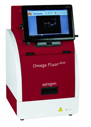
There is a bewildering array of entry level gel documentation systems available these days. Most do exactly what they say on the box in that they can capture images of your gels and blots. So in many cases the customer will just opt for something within their budget and have a high expectation that it will do the job. The Omega Fluor and Omega Fluor Plus from Gel Company/Aplegen will do just that and there is no need to look any further.
However, the Omega Fluor and Omega Fluor Plus are more than just run-of-the-mill gel documentation systems. These systems give you everything you need for exception DNA and protein imaging, plus they are also packed with features that you only expect to see on much higher specification and hence higher price range products....
Syngene, a world-leading manufacturer of image analysis solutions, is pleased to announce its G:BOX Chemi XX6 multi-application imager is being utilised by scientists at the University of Brescia for analysing nitrated proteins.
This is providing the researchers with accurate information on changes in proteins and may help to identify biomarkers related to Alzheimer’s disease. Researchers in the Department of Molecular and Translational Medicine (DMMT) at theUniversity of Brescia in Italy are using a G:BOX Chemi XX6 system to analyse nitrated proteins fluorescently stained with Cy2, Cy3 and Cy5 on 2D DIGE (Difference Gel Electrophoresis) gels. These proteins have been isolated from the...
Prior Scientific has announced that the Core Imaging Facility at the UK's prestigious National Institute for Medical Research (NIMR) has chosen a Prior HLD117 linear motor stage to be the sample platform at the centre of a new system based around an Olympus IX83 microscope.
NIMR is one of the world’s leading medical research institutes. It is dedicated to studying important questions about biological processes that are relevant to all aspects of health. Research at NIMR covers a broad spectrum of basic biomedical science, including infectious diseases, immunology, cell and developmental biology, neuroscience and structural biology. The world-class facilities for research include biological imaging resources, the...
The Imagel Gel Doc system for iPhone or Android is the smart way to quickly grab your gel images. Just hook up your smart phone to the compact hood and start to collect images of all your gels, quickly and easily. The Imagel can be used with 3 different light sources. Use the blue LED transilluminator for DNA applications that use Green DNA, GelGren, SYBR® Green, SYBR® Safe, SYBR® Gold and for Protein applications using LavaPurple....
Cytation™ 5 combines automated digital widefield microscopy with conventional multi-mode microplate detection to provide phenotypic cellular information and well-based quantitative data.
Take advantage of this exciting free Imaging Starter Set offer, which includes:
The new noise-free UC90 microscope camera from Olympus features a large Field-of-View 9 megapixel CCD sensor, creating highly detailed images for on-screen evaluation and discussions with unprecedented clarity.
Maximising the information captured from life and materials science samples is now possible with ease and simplicity, with the new Olympus UC90 microscope camera. Thanks to a range of innovative features, the UC90 excels in capturing and documenting real-life details, while a 4K UHD mode supports scientists in overcoming the limitations of the oculars with the many benefits of complete on-screen operation. In-depth sample...
Introducing ZEISS MultiSEM 506 acquiring more than 2 Tera pixel per hour
At the annual Neuroscience meeting in Chicago, October 17- 21, 2015, ZEISS presented a new variant of the world’s fastest scanning electron microscope: ZEISS MultiSEM 506 features 91 beams working in parallel and increases the throughput of the ZEISS MultiSEM 505 by a factor of three. The unrivaled net acquisition speed of more than 2 Tera pixel per hour enables large-scaled experiments such as imaging of cubic millimeters of brain tissue at nanometer resolution for the analysis of...
Michigan researchers use Raman spectroscopy to study various childhood diseases
Renishaw, a world leader in metrology and spectroscopy, reports on the use of its inVia confocal Raman microscope in the study of various diseases–with a major focus on childhood diseases–at the Children's Hospital of Michigan and Wayne State University. Dr Michael Klein is a surgeon with the Children's Hospital of Michigan–a part of the Detroit Medical Center (DMC)–and a researcher with the Department of Surgery at Wayne...


 CAMECA, a unit of AMETEK Materials Analysis, was recognized among the winners of the 54th Annual R&D 100 Awards by the editors of R&D magazine for its development of the LEAP 5000. Launched in August 2014, the LEAP 5000 is the latest generation of atom probe microscopes, which offer precise atom-by-atom identification, 3-D spatial positioning, and accurate atomic-scale reconstruction of a material’s microstructure...
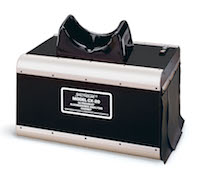 One such product is Spectroline
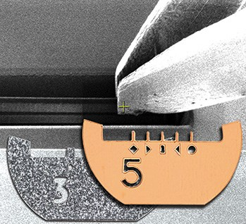 The new EM-Tec range of FIB lift-out grids from EM Resolutions provides a secure way to attach TEM lamella prepared with Focussed Ion Beam (FIB) instruments. Available in multiple post configurations, with a shape optimized for easy accessibility, EM-Tec FIB grids are compatible with all standard 3 mm TEM grid holders.
 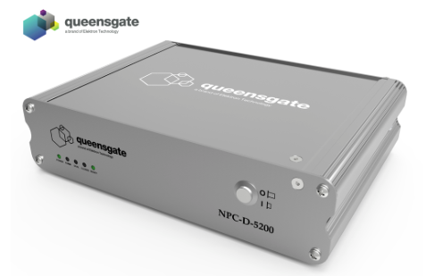 Queensgate pioneered the use of capacitive sensors to provide precise positional feedback in closed loop; the NPC-D-5200 incorporates a precision capacitive measurement circuit and is updated with the stage position 120000 times per second, delivering high positional accuracy at speed. It has the capacity to address a broad spectrum of highly demanding alignment and metrology applications. By employing proprietary low noise...
 Ovizio Imaging Systems, an innovative Belgian company developing cell counting solutions based on quantitative microscopy for life sciences applications, today announces it has secured a funding round of €8m ($9.1m) co-led by New Science Ventures, a US-based venture capital firm, and a private investor. This funding round...
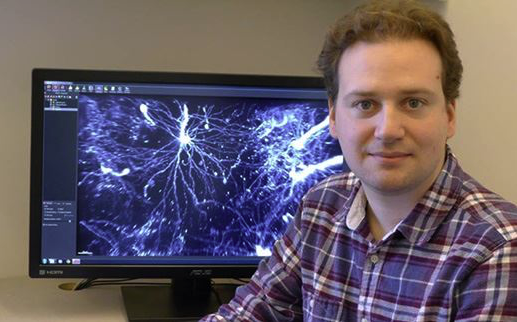
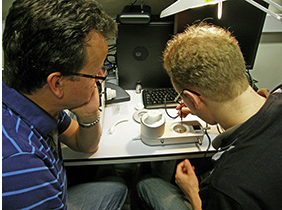 Dr Paul Verkade is a Reader in Cell Imaging in the School of Biochemistry at the University of Bristol where he also heads the Electron Microscopy unit of the Wolfson Bioimaging Facility. His current research focus is to develop techniques and tools for the use of Correlative Light Electron Microscopy (CLEM) studying endocytic sorting. Dr. Verkade is currently chair of the Electron Microscopy section of the Royal Microscopy Society and chair of...
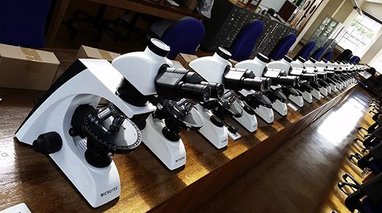 The microscopes have already been used in the undergraduate teaching laboratories by students returning for the new academic year. The School of Earth and Ocean Sciences at Cardiff University offers students a research-led experience across a wide raft of disciplines. They provide undergraduates with top class facilities that include dedicated IT labs, state-of-the-art analytical equipment and even their own research vessel. To coincide...
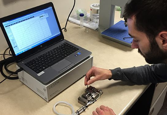 London where it is used to measure load displacement curves on nanocellulose-reinforced polymer composites. Dr Koon-Yang Lee is a Lecturer in Composite Manufacturing in the Department of Aeronautics at Imperial College London. His young and dynamic research group focuses on the manufacturing of polymer (nano)composites, surface and interface engineering, particle stabilised emulsions and foams. His research is highly...
 NIMR is one of the world’s leading medical research institutes. It is dedicated to studying important questions about biological processes that are relevant to all aspects of health. Research at NIMR covers a broad spectrum of basic biomedical science, including infectious diseases, immunology, cell and developmental biology, neuroscience and structural biology. The world-class facilities for research include biological imaging resources, the...
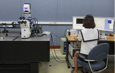 Established in 1976 for R&D of chemical technology in Korea, the Korean Research Institute of Chemical Technology (KRICT) has helped drive the growth of the country“s chemical industry. The focus is on the development of world-class key technologies. There are four key research fields: the development of eco-friendly chemical process technology; the development of high value-added green chemical materials; the discovery of...
 The financial terms of the agreement have not been disclosed. Ovizio has collaborated with Pall for several years to customize the iLine S microscope and will now further improve the technology. The microscope has been specifically designed for use with Pall’s technology and will be commercialized as part of Pall’s Xpansion® cell production platform; the first fully-closed bioreactor for large-scale production of adherent stem cells. The partnership...
 However, the Omega Fluor and Omega Fluor Plus are more than just run-of-the-mill gel documentation systems. These systems give you everything you need for exception DNA and protein imaging, plus they are also packed with features that you only expect to see on much higher specification and hence higher price range products....
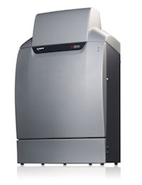 This is providing the researchers with accurate information on changes in proteins and may help to identify biomarkers related to Alzheimer’s disease. Researchers in the Department of Molecular and Translational Medicine (DMMT) at theUniversity of Brescia in Italy are using a G:BOX Chemi XX6 system to analyse nitrated proteins fluorescently stained with Cy2, Cy3 and Cy5 on 2D DIGE (Difference Gel Electrophoresis) gels. These proteins have been isolated from the...
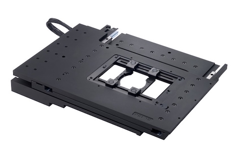 NIMR is one of the world’s leading medical research institutes. It is dedicated to studying important questions about biological processes that are relevant to all aspects of health. Research at NIMR covers a broad spectrum of basic biomedical science, including infectious diseases, immunology, cell and developmental biology, neuroscience and structural biology. The world-class facilities for research include biological imaging resources, the...
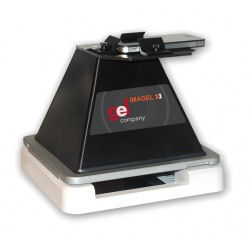 The Imagel Gel Doc system for iPhone or Android is the smart way to quickly grab your gel images. Just hook up your smart phone to the compact hood and start to collect images of all your gels, quickly and easily. The Imagel can be used with 3 different light sources. Use the blue LED transilluminator for DNA applications that use Green DNA, GelGren, SYBR® Green, SYBR® Safe, SYBR® Gold and for Protein applications using LavaPurple....
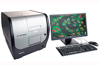 Take advantage of this exciting free Imaging Starter Set offer, which includes:
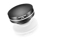 Maximising the information captured from life and materials science samples is now possible with ease and simplicity, with the new Olympus UC90 microscope camera. Thanks to a range of innovative features, the UC90 excels in capturing and documenting real-life details, while a 4K UHD mode supports scientists in overcoming the limitations of the oculars with the many benefits of complete on-screen operation. In-depth sample...
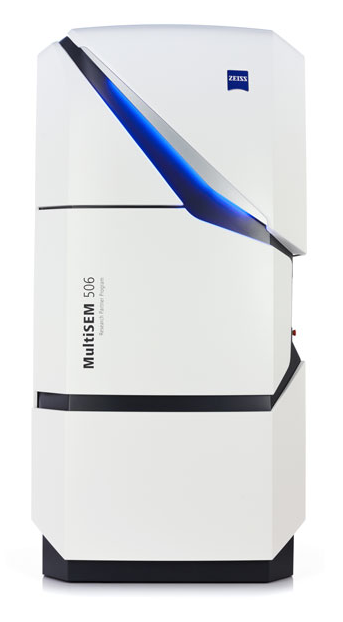 At the annual Neuroscience meeting in Chicago, October 17- 21, 2015, ZEISS presented a new variant of the world’s fastest scanning electron microscope: ZEISS MultiSEM 506 features 91 beams working in parallel and increases the throughput of the ZEISS MultiSEM 505 by a factor of three. The unrivaled net acquisition speed of more than 2 Tera pixel per hour enables large-scaled experiments such as imaging of cubic millimeters of brain tissue at nanometer resolution for the analysis of...
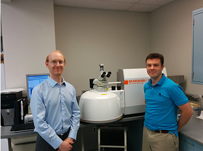 Renishaw, a world leader in metrology and spectroscopy, reports on the use of its inVia confocal Raman microscope in the study of various diseases–with a major focus on childhood diseases–at the Children's Hospital of Michigan and Wayne State University. Dr Michael Klein is a surgeon with the Children's Hospital of Michigan–a part of the Detroit Medical Center (DMC)–and a researcher with the Department of Surgery at Wayne...